REVIEW ARTICLE
MOREIRA, Danilo José Silva [1], FONSECA, Juliana Brito da [2], ROSSI, Karoline [3], VASCONCELOS, Suzana dos Santos [4], OLIVEIRA, Vinicius Faustino Lima de [5], DIAS, Claudio Alberto Gellis de Mattos [6], OLIVEIRA, Euzébio de [7], DENDASCK, Carla Viana [8], ARAÚJO, Maria Helena Mendonça de [9], BRITO, Maysa Vasconcelos [10], FECURY, Amanda Alves [11]
MOREIRA, Danilo José Silva. Et al. General aspects of xeroderma pigmentosum: A review. Revista Científica Multidisciplinar Núcleo do Conhecimento. Year 05, Ed. 03, Vol. 11, pp. 114-126. March 2020. ISSN: 2448-0959, Access Link: https://www.nucleodoconhecimento.com.br/health/xeroderma-pigmentosum , DOI: 10.32749/nucleodoconhecimento.com.br/health/xeroderma-pigmentosum
SUMMARY
Xeroderma Pigmentoso (XP) is a rare, recessive and autosomal genetic disease that also affects both sexes and all ethnicities, being closely associated with communities with a high rate of inbreeding. The aim of this review was to detail the main routes of DNA repair of XP, the different functional defects that result in the development of the 8 types of XP, the main characteristics of the clinical picture of a patient with XP, the main comorbidities associated with XP, and the treatments available or that are still in studies for individuals affected by XP. The bibliographic research was carried out in the databases: Redalyc, Institutional Repository of the Federal University of Juiz de Fora, Scielo, Brazilian Digital Library of Theses and Dissertations, Science Research.com, Lilacs and Pub Med, using keywords or their associations: Xeroderma – Xeroderma Pigmentoso. XP is a genetic disease that has no cure; the individual with XP has a photosensitive skin and, when exposed to UV radiation, may develop several dermatological complications; the manifestations of XP are directly linked to the genetic defect; NER is undoubtedly the main route of DNA repair when it comes to XP; in XP-V the by-pass of the tape with the DNA lesion is not done by polymerase pol eta but by another polymerase of the Family Y; defects in DNA repair pathways can cause not only XP, but also other diseases; and the treatment for XP is palliative. It consists of the use of specific UV protectors, drugs, repair enzymes and adenoviral vectors, as well as cryosurgery, photodynamic therapy (PDT), surgical removal of tumors and psychological follow-up.
Keywords: Xeroderma Pigmentoso, repair, comorbidity, treatment.
INTRODUCTION
Xeroderma Pigmentoso (XP) is a rare, recessive and autosomal genetic disease that also affects both sexes and all ethnicities, being closely associated with communities with a high rate of inbreeding (DANTAS, 2018; OLIVEIRA, 2003).
It is known that there is a relationship between the development of XP and defects in DNA repair, which consist of mechanisms used by the cell to correct dna damage, avoiding the induction of mutations (SANTIAGO, 2015).
The development of each of the eight types of XP is conditioned by different functional defects, which involve DNA repair pathways (SANTIAGO, 2015).
Individuals with XP, as well as in any other disease, present symptomatological characteristics that lead to the establishment of a clinical picture, which corresponds to the set of symptoms presented by the patient (PRIBERAM DICIONÁRIO, 2020).
In the literature, there is evidence of the association between XP and the development of other comorbidities, which consist of the occurrence of two or more diseases at the same time in an individual (GOLDMAN; AUSIELLO, 2011).
Over time, to improve the quality of life of an individual with XP, several treatments have been developed, which consist of ways to care for a patient (SCOTTINI, 2017).
GOALS
Detail the main ways of repairing XP’s DNA.
Detail the different functional defects that result in the development of the 8 types of XP.
Detail the main characteristics of the clinical picture of an individual with XP.
Detail the main comorbidities associated with XP
Detail available treatments or treatments that are still in studies for individuals affected by XP.
METHOD
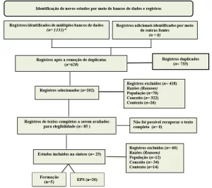
The bibliographic research was carried out in the databases: Redalyc, Institutional Repository of the Federal University of Juiz de Fora, Scielo, Brazilian Digital Library of Theses and Dissertations, Science Research.com, Lilacs and Pub Med, using keywords or their associations: Xeroderma – Xeroderma Pigmentoso.
The inclusion criteria used in the search were the comprehensive online availability, the direct approach on XP or on some relevant aspect in relation to this pathology and text of the work written in English, Portuguese or Spanish. In relation to the exclusion criteria applied, there are duplicate works and work carried out before the year 2000.
A search was carried out in the databases mentioned in order to list the existing studies related to XP in the literature. From a previous reading of the titles and abstracts of the papers found, productions that did not meet what was expected to write this review were discarded. The full text of the productions that went through the previous stages was then read, selecting for this review those that addressed relevant aspects of XP.
RESULTS
DNA AND XP REPAIR PATHWAYS
The variant form of XP (XP-V) is linked to the absence of polymerase pol eta present in the transinjury repair pathway (TLS) (CASTRO, 2016). All other types of XP manifestations are directly linked to defects in the Nucleotide Excision Repair (NER) pathway, which is formed by the joint action of about 30 proteins (SANTIAGO, 2015).
NER recognizes spatial distortions in the structure of the DNA molecule, correcting lesions caused by the action of ultraviolet (UV) light, products derived from the activation of polycyclic aromatic hydrocarbons, oxidative products and products formed by chemotherapy (MORI, 2015; MOURA, 2015; SANTIAGO, 2015).
NER has two pathways: Transcription Coupled Repair (TCR) and Genome Global Repair (GGR) (MORI, 2015; MOURA, 2015; SANTIAGO, 2015).
TLS makes a by-pass of a DNA spatial lesion and the main polymerase involved in this process is the pol eta. In the absence of pol eta, the polymerase that makes the by-pass of the lesion is the pol iota, which has a much higher error rate than pol eta (CASTRO, 2016; LERNER, 2014).
GENETIC MECHANISMS INVOLVED WITH THE DEVELOPMENT OF XP
The seven proteins of the Xeroderma Pigmentoso complementation group – XPA, XPB, XPC, XPD, XPE and XPF and XPG – are part of the NER pathway of DNA. Complications in the genes encoding these proteins result in XP due to NER inefficiency (MORENO, 2017; SANTIAGO, 2015).
In XP-V, the functioning of the NER pathway of DNA is normal, however, the function of polymerase pol η, an enzyme synthesized from the POLH gene and responsible for DNA by-pass, is compromised, causing inefficiency of the TLS pathway (MORENO, 2017).
CLINICAL PICTURE OF XP
Patients with XP may present broad-spectrum clinical manifestations: sensitivity to UV radiation, predisposition to tumor development, premature aging of the skin, erythemas, neurological complications and impaired body development (CHAIBUB, 2011; DANTAS, 2018; LEITE, 2008; VIANA, 2011).
COMORBIDITIES ASSOCIATED WITH XP
Cockayne Syndrome, Trichothiodystrophy and XP/deSanctis-Cacchione Syndrome are diseases that are associated with XP due to defects that make it impossible for DNA repair pathways to function perfectly. This syndrome also include sensorineural hearing loss, cataract, pigmentary retinopathy, skin photosensitivity and dental caries. (CASTRO, 2016; SOLTYS, 2010).
In Trichothiodystrophy, the affected individual may present with short stature, microcephaly, cognitive deficit, characteristic facy – retreated chin and large ears – nail dysplasia, photosensitivity, ichthyosis, inferectility and greater predisposition to infectious diseases (CASTRO, 2016; SOLTYS, 2010).
XP/deSanctis-Cacchione syndrome consists of an XP picture associated with the coexistence of hypogonadism, neonism and severe neurological disease in the affected individual (LERNER, 2014; SOLTYS, 2010).
TREATMENTS AVAILABLE
Basically, the treatment revolves around the removal of any tumors that arise and changes in the life habits of the affected individual, such as the use of long clothes and the exercise of activities at night (LERNER, 2014).
Isotretinoin (cisretinoic acid) is used in some cases; another treatment tool is the use of adenoviral vectors (LEITE, 2008). Cryosurgery or cryotherapy is also an alternative tool for the treatment of XP patients (ANTUNES; ANTUNES; SILVA, 2007). The use of PDT is also a tool that allows the treatment of cancers from XP (ZAMARRÓN et al., 2017).
DISCUSSION
NUCLEOTIDE EXCISION REPAIR PATHWAY
As for the NER steps, the lesion is first identified, followed by the opening of the double tape and the verification of the lesion. Soon after, the pre-incisional complex is formed that causes the cutting of the oligopeptide containing the lesion and repair synthesis (MORI, 2015).
Ner’s two sub-routes – TCR and GGR – are classified according to the cellular machinery used to perform the repair (SANTIAGO, 2015).
Basically, TCR-NER occurs when there is a blockage to the progression of RNA polymerase I or II (RNA in). The CSB protein (Cockayne Syndrome Complementation group B) has a high affinity for RNA in and it is believed that this interaction increases in intensity when RNA pol encounters some physical obstacles that prevent the continuity of the transcription process, resulting in an intense interaction of CSB and the transcriptional complex. Therefore, CSB is considered the first protein that responds to an injury in the TCR-NER process (MORI, 2015).
The increased interaction between CSB and RNA in would occur when the transcription was interrupted by some lesion on the mold tape, which is believed to displace CSB backtracking. This displacement would vacate binding sites and open up other repair proteins. According to RNA pol regresses, replication protein A (RPA) binds to the sites of the simple tape preventing renaturation, in addition to signaling the recruitment of XPA to the site of the injury. CSB also recruits CSA (Cockayne syndrome complementation group A). CSA acts in the ubiquinization of CSB, a process that is very important for TCR-NER, since chimeric CSB has the ability to recover any molecular defects in CSB. From that moment on, injury repair mechanisms are common between TCR-NER and GGR-NER (CASTRO, 2016; MORI, 2015).
The GGR-NER subvia, as stated earlier, acts when damage to the double DNA tape distorts the spatial structure, compromising the double helix. The recognition is given by two protein complexes, the UV-DDB which is a heterodmer resulting from the union of DDB1 and DDB2 and another heterodmer XPC-RAD23B/hHr23B-Centrina 2, which is the first to recognize the lesion. XPC proteins are the component of its heterodmer that actually recognizes the lesion, as it has a DNA binding affinity and has this increased binding capability the more distorted the spatial lesion of the double helix. The UV-DDB complex reduces the repair time of CPD’s and 6-4 PP’s (photoproducts derived from the interaction between UV rays and the skin) due to a stabilizing function it exerts on NER. XP-E patients have a marked decrease in repair of 6-4 PP’s (CASTRO, 2016; FELTES, 2017; MORI, 2015; PUUMALAINEN, et al., 2015).
After the identification process, the subsequent stages of NER follow a common path in both subpathic routes with the recruitment of TFIIH (Transcription Factor Human II), a multiprotein complex with 10 subunits, in which xpb and XPD proteins are found. These are ATP-dependent helicases that unroll DNA in opposite polarities. The XPD rolls out in the 5′-3′ direction, while the XPB in the 3′-5′ direction. XPB can be more efficient in assembling TFIIH than in the helicase process. XPD has a very narrow arc-shaped structural domain through which it moves through the simple unrolled tape. As the size of the hole formed by the arc is not large, XPD can be considered a damage check factor, as injuries of large extents will not pass due to the physical obstacle. In this context, up to this point there is the opening of the double tape and the verification of the lesion. Following the process, the next step is to assemble the pre-incisional complex, with the connection of XPA, RPA and XPG and shutdown of the XPC-hHR23B complex. One of the functions of XPA is to detect some injury that was not noticed by XPD, as well as connect in the opposite direction to that of XPB, at the 5′ end of the injured tape. The RPA connects the uninjured tape covering about three dozen nucleotides, and to assist in the correct positioning of endonucleases, in addition to displacing the XPC, which leaves the TFIIH complex. XPG and the ERCC1-XPF complex are the endonucleases that received the aid of RPA to position themselves and that will excise the oligomer with the lesion and allow the continuity of the repair process (CASTRO, 2016; MORI, 2015; SANTIAGO, 2015).
To finish the process, a new simple DNA tape is synthesized to occupy the damaged site, with the action of polymerases in δ and pol η in the synthesis of the new tape and the action of DNA ligase I or II, which amends the new tape to the site (CASTRO, 2016; FELTES, 2017).
THE REPAIR ROUTE BY TRANSLESION SYNTHESIS
The existence of a DNA lesion results in a change in the spatial structure of the double tape, making it impossible to recognize the nitrogen bases by the replicative polymerases of family A (α,β,ε), which function perfectly only in intact DNA, which can lead to the stopping of the replication fork and consequently cellular apoptosis (LERNER, 2014; MORENO, 2017).
Polymerases of the Y family, mainly pol eta, are the most qualified to perform the repair of damaged DNA, because they have smaller catalytic palm and thumb sites, which allows the insertion of nucleotides even with the presence of the lesion in the DNA ribbon. With this process, the replication fork is prevented for a long period, which could trigger a process of cellular apoptosis (CASTRO, 2016).
At the moment when replicative polymerases are prevented from replicating by some physical impediment in the DNA tape, the nuclear cell proliferation antigen (PCNA) will have its lysine 164 monoubiquitinized by the ubiquitin complex ligase E2/E3. This change promotes the increased affinity of PCNA with polymerases of the Y family, providing a call for tls polymerases, resulting in the substitution of proliferative polymerases, and interaction of Pol eta with the site of the lesion constituting the by-pass (CASTRO, 2016).
Made the deviation by Pol eta or another polymerase of the Family Y, another polymerase of the family B, mainly Pol zeta, inserts bases for a few more pairs ahead. After the by-pass, Pol eta is desubiquitinized by USP7 protein. With this, polymerases of the Family A, mainly Pol delta, continue the process of DNA replication (CASTRO, 2016; LERNER, 2014).
THE TYPES OF XERODERMA PIGMENTOSO
Xeroderma pigmentosum type A (XP-A) consists of a phenotypic expression derived from alteration in the XPA gene, which has 6 exons located on chromosome 9q22.3 and extension of 25 kpb. The protein transcribed from the XPA gene has 42 kDa and domains that support its binding to DNA, Zinc Fingers (DANTAS, 2018; LEITE, 2008). Patients with deficiency in this cellular mechanism have impaired the ability to properly form the pre-incisional complex, which would be the correct arrangement of cellular machinery at the ends of dna injury (BENSENOUCI et al., 2016). In Japan and Asia, most cases of XP are related to the XPA gene, presenting mutation in the last nucleotide of exon 3 (LEITE, 2008).
Xeroderma Pigmentosum type B is a form of manifestation of the disease resulting from the non-synthesis of the XPB/ERCC3 Protein. The XPB gene is located on chromosome 2q21 and its deficiency harms the nervous system, as well as is part of Cockayne syndrome (DANTAS, 2018). The XPB protein is an ATP helicase (Adenosine Triphosphate) dependent that follows the DNA tape in the 3′-5′ direction, recruited after initial recognition of the lesion at the time the TCR (Transcription Coupled Repair) and GGR (Global Genomic Repair) converge to a common NER pathway (CASTRO, 2016).
Xeroderma Pigmentosum type C (XP-C), in turn, occurs when genetic abnormalities affect the XPC gene, which has 16 exons and 33 Kpb and is located in the 3p25 locus. The protein transcribed from the XPC gene has 940 amino acids and binds to other proteins, hHR23B and centrine 2, forming a protein complex with terminal C-binding domain to human DNA and transcription factor II, which recognizes DNA lesions (LEITE, 2008). The most common mutation in the XPC gene is a 2bp deletion. According to an Algerian study analyzing 17 patients with XP-C, all had homozygosis for 2bp deletion. The most frequent deletion results in an inefficient termination codon, impairing the interaction between proteins XPC, hHR23B, Centrine 2 and TFIIH, which are essential in the recognition of DNA damage, and the beginning of the NER repair pathway (BENSENOUCI et al., 2016).
Xeroderma Pigmentosum type D is associated with defects in the XPD gene, which is located on chromosome 19.q13.2-13.3. The protein transcribed from this gene is related to excision and DNA repair. Defects in the expression of this protein are also associated with diseases such as Trichodystrophy and Cockayne Syndrome (DANTAS,2018). The XPD protein is a DNA helicase that is part of TFIIH, responsible for stabilizing the proteins at the site of the lesion. In addition, XPD is ATP dependent, and unwinds the double DNA tape in the 5′-3′ direction, the inverse of the XPB protein (CASTRO, 2016; SOLTYS, 2010).
Xeroderma Pigmentosum type E (XP-E) is linked to mutations on chromosome 11p12-p11. The XPE gene is responsible for dna-bonding protein synthesis (DANTAS, 2018). The product encoded from the XPE gene is the DDB2 protein (FELTES, 2017). The XPE protein is formed by subunits p127/DDB1 and p48/DDB2 and participates in the pathways of correction of DNA lesions, recognizing lesions that were not recognized by the XPC-hHR23B complex as cyclobutane pyrimidine dyes (CPD’s) (LERNER, 2014). XPE is used only in the GGR-NER sub- route. In the case of TCR, both XPE protein and XPC-hHR23B are not required. The physical blockade of DNA for the continuation of RNA polymerase II is the sign of recruitment of this protein (LEITE, 2008).
Xeroderma Pigmentosum type F (XP-F) comes from a mutation in the gene located on chromosome 16p13.3-p13 (DANTAS, 2018). The XPF protein binds to the ERCC1 protein and forms the heterodmer XPF-ERCC1. The XPF protein is a product of the XPF/ERCC4 gene, with 103 kDa. The stabilization of the complex is associated with the union of the two proteins. The XPF-ERCC1 complex has a domain located in XPF with catalytic nucleasse properties, participating in the NER pathway of DNA. It is also present in other DNA repair pathways, such as DNA DSB/QFD (Double Strand break) and cross-linking between DNA tapes (LERNER, 2014). The incision of the oligonucleotide that possesses the lesion only occurs after the arrival of the XPF-ERCC1 complex, which is the first to make the incision in the 3′ direction, leaving a hydroxyl radical in which the repair synthesis (MORI, 2015) will be initiated by a DNA polymerase enzyme.
Xeroderma Pigmentosum type G (XP-G) is associated with abnormalities in the gene located on chromosome 13q33 (DANTAS,2018). The XPG protein is an endonuclease that breaks down the injured DNA, and its action begins about 5 nucleotides before the lesion site in the 3′ direction. The incision in the direction 5′ by heterodmer XPF/ERCC1 also requires the presence of XPG protein in the NER repair pathway. Patients who manifest XP-G disease have extremely deficient NER, and deficiencies in the gene encoding XPG result in the XP/CS phenotype, when the individual concomitantly presents Xeroderma pigmentosum and Cockayne Syndrome. XPG acts as a PC4/CF4 transcription coactivator (Positive Cofactor 4 or Positive Cofactor 4) that prevents mutation and cell death from oxidative DNA damage. PC4 works by removing XPG from regions where RNA polymerase has found an injury, releasing the site for the repair process. In addition, XPG stabilizes TFIIH and allows phosphorylation of nuclear receptors, having an important role in gene expression (SOLTYS, 2010).
Variant Xeroderma Pigmentosum (XP-V) is a disease resulting from the absence of polymerase in η, encoded from the POLH gene. The η is composed of 11 éxons, 713 amino acids and its central function is to perform dna by-pass (MORENO, 2017). XP-V patients have the dna repair pathway by normal nucleotide excision, but have other defective repair mechanisms, such as the transfunction mechanism that in addition to metabolizing photo products, also degrades reactive oxygen species (LIU; CHEN, 2006). The η can replicate DNA with lesions by making deviations, decreasing the incidence of cancer by decreasing mutagenesis. In XP-V patients, sites injured due to UV light exposure have dna replication interrupted and the course of the process is more prone to errors because pol η is the most specialized polymerase in by-pass. In the absence of pol η, other polymerases of the same family (such as pol ι) make the deviation, but with low specificity of the insertion of corresponding bases in the complementary tape, causing numerous mutations that result in skin cancers (LERNER, 2014; MORENO, 2017).
Polymerase pol η can perform the by-pass in the two main photoproducts formed after exposure to UV light – cyclobutane pyrimidine dyes and 6.4 PP’s – and also act on oxidized bases (MORENO, 2017). The knowledge that is acquired until 2017 about XP-V is that its development takes place from contact with UVB rays and UVC. However, this relationship cannot be delimited only with these wavelengths, since there are few studies describing the mutagenic effects of UVA in XP-V patients (MORENO, 2017). In the State of Goiás, located in Brazil, a community in the city of Araras has a frequency of one case of XP-V per 200 people (MORENO, 2017).
THE CLINICAL MANIFESTATIONS OF XERODERMA PIGMENTOSO
Individuals with XP-A follow a clinical course that results in the onset of neurological symptoms already in childhood. In patients with XP-A who have reached adulthood, extensive neuronal loss and gliosis of the white matter in the central nervous system (CNS) is observed. It is believed that acquired factors, such as oxidative stress or random toxicity of amino acids, may be related to neurological manifestations in patients in XP-A (UEDA et al., 2012).
Regarding XP-C, it was found by missionaries from Good Samaritans International in partnership with Guatemalan physicians that a guatemalan village has a high incidence of XP-C patients. There was no presentation of neurological symptoms, however the children were no more than ten years old, had advanced tumor proliferation, gangrene and infection, with reports of severe pain after exposure to the sun (CLEAVER, et al., 2011).
Due to the important function of the XPD protein within the NER repair pathway, XP-D patients present severe NER pathway deficiency, leading to neurological degeneration of homozygous patients in about 20% to 30% of cases (RIBEIRO et al., 2018).
Patients with XP-E, similar to XP-C patients, usually do not have neurological diseases, although they may present cns abnormalities due to genetic and environmental reasons in some cases (ORTEGA-RECALDE et al., 2013).
Of all patients with XP, 20% are XP-V and have a more favorable prognosis than classic XP (XP-A to XP-G), deficient in NER. The photosensitivity of patients with V-XP is accentuated in early adulthood, when the first malignant lesions appear. In addition, individuals with XP-V do not present neurological lesions (LERNER, 2014).
Neurological manifestations are present in complementation groups XP-A, XP-B, XP-D and XP-G (BENSENOUCI et al., 2016).
XP individuals are more likely to develop oculocutaneous neoplasms in relation to the others, with the main types being basal cell carcinoma (BCC), squamous cell carcinoma (SCC) and melanoma (FELTES, 2014; HALKUD et al., 2014; OLIVEIRA et al., 2003).
As for ocular neoplasms, eyelids, conjunctiva and cornea are the regions most susceptible to the development of neoplasms (HALKUD et al., 2014).
In the literature, there are also reports that regions such as the oral cavity, tip of the tongue, bone marrow, testicles, stomach, lungs and pancreas in XP patients are more likely to be affected by a neoplasm compared to other people (FELTES, 2017; HALKUD et al., 2014).
THE RELATIONSHIP BETWEEN XERODERMA PIGMENTOSO AND THE DEVELOPMENT OF COMORBIDITIES
The appearance of defects in the NER pathway of DNA susceptibilities the affected individual to develop not only classic XP, but also other autosomal and recessive diseases, such as Cockayne Syndrome, Trichotriodystrophy, Brain-Oculus-Skeletal Syndrome, UV-sensitive syndrome, XPF-ERCC1 Syndrome and XP/deSanctis-Cacchione Syndrome, both presenting as a common feature the increase in sensitivity to UV rays (MOURA, 2015; SOLTYS, 2010).
Cockayne Syndrome is a clinical condition associated, in most cases, with neurological and physical development problems, being classified into five complementation groups according to the affected gene locus. In two of these groups, mutations occur in the CSA (ERCC8) and CSB (ERCC6) genes, resulting in a phenotype with the Impaired DNA TCR pathway. In the other groups, mutations affect the XPB (ERCC3), XPD (ERCC2) or XPG (ERCC5) genes, which, in turn, result in a phenotype with the ner pathway deficient. As a result, the NER-deficient groups correspond to individuals who are susceptible to developing both Cockayne Syndrome and XP, with higher chances of developing skin cancer (CASTRO, 2016; SOLTYS, 2010). The clinical picture of the individual with Cockayne Syndrome may also include sensorineural hearing loss, cataract, pigmentary retinopathy, skin photosensitivity and dental caries (CASTRO, 2016).
Trichothiodystrophy is a condition in which the affected individual does not properly perform the formation of sulfide bridges, thus interfering in the synthesis of some amino acids that make up the hair and nails, such as cysteine. This explains a peculiar characteristic of the disease: fragile nails and brittle hair and drains (CASTRO, 2016; SOLTYS, 2010). Other manifestations that may appear in the clinical picture of an individual with Trichothiodystrophy include short stature, microcephaly, cognitive deficit, characteristic facy – retreated chin and large ears – nail dysplasia, photosensitivity, ichthyosis, infertility and greater predisposition to infectious diseases (CASTRO, 2016). Regarding the genetics of the disease, four genes associated with the development of Trichothiodystrophy have already been identified: XPD, XPB, TTDA and TTDN1. While the latter does not have its functions clarified, it is known that the first three encode proteins that integrate TFIIH, a structure associated with DNA NER pathway (SOLTYS, 2010).
XP/deSanctis-Cacchione Syndrome consists of an XP picture associated with the coexistence of hypogonadism, neonism and severe neurological disease in the affected individual. The scholars who named the syndrome were the first to establish a relationship between XP and neurological abnormality (LERNER, 2014; SOLTYS, 2010).
In addition to the diseases mentioned, it is also observed that XP individuals are more likely to develop oculocutaneous neoplasms in relation to the others, a risk that is evaluated at a thousand times higher. In cases of type C and E XP, there is a higher incidence of skin cancer (FELTES, 2014; HALKUD et al., 2014; OLIVEIRA et al., 2003).
The main types of oculocutaneous neoplasms affecting XP individuals are basal cell carcinoma (BCC), squamous cell carcinoma (SCC) and melanoma, the first two being the most frequent. BCC is characterized by slower growth and lower chances of metastatizing, while CPB has opposite characteristics (HALKUD et al., 2014; OLIVEIRA et al., 2003).
As for ocular neoplasms, the higher exposure to UV rays of the sun mainly susceptibilities eyelids, conjunctiva and cornea to the development of these neoplasms.
In the literature, there are reports that regions such as the oral cavity, tip of the tongue, bone marrow, testicles, stomach, lungs and pancreas in XP patients are more likely to be affected by a neoplasm compared to other people (FELTES, 2017; HALKUD, 2014). With higher chances of developing neoplasms, mainly involving the skin, XP patients minimize their life expectancy (MOURA, 2015).
TREATMENT
XP has no cure because it is a genetic disease and the measures adopted for treatment are palliative, seeking to increase the patient’s life expectancy. Basically, the treatment revolves around the removal of any tumors that arise, recommending a follow-up with dermatologist. It is necessary that the XP patient change their life habits, wearing long clothes and decreasing the areas exposed to light. In addition, the exercise of activities in the night, instead of the day and evening period, minimizes the number of the PATIENT XP with the largest source of UV light that is on Earth, the sun (LERNER, 2014).
It is reported that the use of isotretinoin (cisretinoic acid) reduces the onset and proliferation of skin neoplasms in patients with a high incidence of cancer. Another option is the use of repair enzymes encapsulated in liposomes, such as T4 endonucleosis V and CPD-photoliasis, which repair CPD’s lesions. The enzyme T4 endonucleosis V cuts the damaged DNA tape and metabolizes, generating products accessible to cellular machinery. While the CPD photoliasis, breaks bonds in the cyclobutane ring formed between pyrimidins. For the repair of 6-4 PP’s there are the 6.4 PP’s photoliases that need to be activated by the use of UVA radiation, which generates a certain fear in the use of this feature because UVA can cause other injuries that may be problematic to humans (LEITE, 2008).
Another treatment tool is the use of adenoviral vectors. In summary, the terminal domain of globose format of the adenovirus interacts with the CAR receptor (Coxsackie-Adenovirus Receptor). After interactions between the virus and the cell surface, the signal for endocytosis occurs and the adenovirus is internalized, released by the endomo and moved to the cell nucleus by microtubulins. It is necessary to clarify that in the construction of adenoviral vectors, regions are removed and for other of interest to be inserted. Recombinant adenovirus genetic engineering was obtained by means of recombinant genetic engineering for the XPA, XPC, XPD and XPV genes, which are efficient for the transduction and complementation of cellular processes in humans (LEITE, 2008). Through a retrovirus-based technique, XPC protein transcomplementation has been reported, transducing XPC into human stem cells, restoring total NER capacity. Despite in vitro success, in practice retroviruses can cause side effects not controllable by those who apply the technique – such as blood hematopoietic pathways impairments (DUPUY, et al., 2013).
Cryosurgery or cryotherapy is also an alternative tool for the treatment of XP patients, a technique that consists of freezing the injured area and the indication for this type of procedure takes into account the macroscopic aspect of the lesion, dimensions, location, histological composition and age of the patient. Cryosurgery mainly uses liquid nitrogen and consists of a very fast and severe freezing, with subsequent slow thawing, causing cell death of the affected region (ANTUNES; ANTUNES; SILVA, 2007).
The use of photodynamic therapy (PDT) is a tool that allows the treatment of cancers from XP, without invasive methods or use of toxic chemotherapy to healthy tissues. It is reported that clinical studies using methyl-δ-aminolevulinic acid and red light have been shown to be a good alternative for the treatment of superficial basal cell cancer in XP patients (ZAMARRÓN et al., 2017).
CONCLUSION
XP is a genetic disease that has no cure, resulting from defects arising from the NER pathway and in the TLS pathway of DNA repair, and may assume 8 different types of manifestations.
The individual with XP has a photosensitive skin and when exposed to UV radiation can develop several dermatological complications, having an exponentially higher chance of developing basal cell, spirocellular and melanoma cancers when compared to a normal individual for XP, in addition to neurological and ophthalmologic impairments.
Manifestations of XP are directly linked to the genetic defect that precedes its phenotypic manifestation, presenting inefficient points in the DNA repair chain that change according to the type of XP.
NER is undoubtedly the main route of DNA repair when it comes to XP, because 7 types of XP are due to this mechanism of correction of DNA lesions. NER is subdivided into two sub-pathways: TCR-NER and GGR-NER, the first being active in the situations of rna elongation blockand the second when there is spatial damage to the double DNA tape.
In XP-V the by-pass of the tape with dna injury is not done by polymerase pol eta but by another polymerase of the familiaY which results in a much higher rate of errors in the TLS repair pathway. Pol eta can insert nucleotides into the DNA ribbon even with the presence of the lesion, and it is the polymerase of the Y family with the lowest error rate.
Defects in DNA repair pathways can cause diseases other than XP such as Cockayne Syndrome and Trichotriodystrophy, and may even have concomitant manifestations such as XP/deSanctis-Cacchione Syndrome.
Treatment for XP is palliative. It consists of specific UV protectors, drugs, repair enzymes, adenoviral vectors, cryosurgery, PDT, surgical removal of tumors and psychological follow-up.
REFERENCES
ANTUNES, A. A.; ANTUNES, A. P.; SILVA, P. V. A criocirurgia como tratamento alternativo do xeroderma pigmentoso. Revista Odonto Ciência, v. 22, n. 57, p. 228-232, set. 2007.
BENSENOUCI, S.; LOUHIBI, L.; VERNEUIL, H.; MAHMOUDI, K.; SAIDI-MEHTAR, N. S. Diagnosis of Xeroderma Pigmentosum Groups A and C by Detection of Two Prevalent Mutations in West Algerian Population: A Rapid Genotyping Tool for the Frequent XPC Mutation. BioMed Research International, v. 2016, n. 2180946 jun. 2016.
CASTRO, L. P. Caracterização genotípica de pacientes brasileiros com deficiência em processos de reparo de DNA. Tese (Doutorado em Biotecnologia) – Universidade de São Paulo. São Paulo, p. 36. 2016.
GOLDMAN, L.; AUSIELLO, D. Cecil Medicina. 23. ed. Rio de Janeiro: Elsevier, 2011.
CHAIBUB, S. C. W. Alta incidência de Xeroderma Pigmentosum em comunidade no interior de Goiás. Surg Cosmet Dermatol, Goiânia, v. 3, n. 1, p. 81-83, jan/mar. 2011.
CLEAVER, J. E.; FEENEY, L.; TANG, J. Y.; TUTTLE, P. Xeroderma Pigmentosum Group C in an Isolated Region of Guatemala. Journal of investigative dermatology, v.127, n. 2, p. 493-496., fev. 2007.
DANTAS, E. B. XERODERMA PIGMENTOSO: RELATO DE CASO. Tese (Trabalho de Conclusão de Curso de Medicina) – Universidade Federal de Sergipe. Lagarto, p. 38, 2018.
DUPUY, A.; VALTON, J.; LEDUC, S.; ARMIER, J.; GALETTO, R.; GOUBLE, A.; LEBUHOTEL, C.; STARY, A.; PAQUES, F.; DUCHATEAU, P.; SARASIN, A.; DABOUSSI, F. Targeted Gene Therapy of Xeroderma Pigmentosum Cells Using Meganuclease and TALEN. PLoS One, v. 8, n. 11., páginas, nov. 2013.
FELTES, B. C. Estudo conformacional do complexo proteico DDB2-DDB1 e suas diferentes variantes mutantes na doença Xeroderma Pigmentosum. Tese (Doutorado em Biologia celular e molecular) – Universidade Federal do Rio Grande do Sul. Porto Alegre, p. 100. 2017.
HALKUD, R.; SHENOY, A. M.; NAIK, S. M.; CHAVAN, P.; SIDAPPA, K. T.; BISWAS, S. Xeroderma pigmentosum: clinicopathological review of the multiple oculocutaneous malignancies and complications. Indian Journal of Surgical Oncology, v. 5, n. 2, p. 120-124, abr. 2014.
LEITE, R. A. Uso de vetores adenovirais no diagnóstico de portadores de xeroderma pigmentosum e em estudos de reparo de DNA. Tese (Doutorado em microbiologia) – Universidade de São Paulo. São Paulo, p. 41, 2008.
LERNER, L. K. Papel das proteínas XPD e DNA polimerase eta nas respostas de células humanas a danos no genoma. Tese (Doutorado em Ciências) – Universidade de São Paulo. São Paulo, p. 201. 2014.
LIU, G.; CHEN, X. DNA Polymerase η, the Product of the Xeroderma Pigmentosum Variant Gene and a Target of p53, Modulates the DNA Damage Checkpoint and p53 Activation. American Society of Microbiology, v. 26, n. 4, p. 1398-1413, fev. 2006.
MORENO, N. C. Efeitos da luz UVA em células de pacientes com Xeroderma Pigmentosum Variante. Tese (Doutorado em Genética) – Universidade de São Paulo. São Paulo, p. 39. 2017.
MORI, M. P., Novo papel da proteína XPC na regulação dos complexos da cadeia de transporte de elétrons e desequilíbrio redox. Tese (Doutorado em Bioquímica) – Universidade de São Paulo. São Paulo, p. 174. 2015.
MOURA, L. M. S. Busca de variantes em sequência de DNA proveniente de pacientes com deficiência em processos de reparo do genoma. Tese (Mestrado em Bioinformática) – Universidade de São Paulo. São Paulo, p. 80. 2015.
OLIVEIRA, C. R. D; ELIAS, L.; BARROS, A. C. M; CONCEIÇÃO, D. B. Anestesia em Paciente com Xeroderma Pigmentoso. Relato de Caso. Revista Brasileira de Anestesiologia, Florianópolis, v. 53, n. 1, p. 46-51, jan/fev. 2003.
ORTEGA-RECALDE, O. O.; VERGARA, J. I.; FONSECA, D. J.; RIOS, X.; MOSQUERA, H.; BERMUDEZ, O. M.; MEDINA, C. L.; VARGAS, C. I.; PALLARES, A. E.; RESTREPO, C. M.; LAISSUE. P. Whole-Exome Sequencing Enables Rapid Determination of Xeroderma Pigmentosum Molecular Etiology. Plos One, v. 8, n. 6, jun. 2013.
PRIBERAM DICIONÁRIO. Disponível em:<https://dicionario.priberam.org/quadro%20clinico>. Acesso em: 24 mar. 2020.
PUUMALAINEN, M. R.; RUTHERMANN, P.; MIN, J. H.; NAEGELI, H. Xeroderma pigmentosum group C sensor: unprecedented recognition strategy and tight spatiotemporal regulation. Cellular and Molecular Life Sciences, v. 73, 547-566, out. 2016.
RIBEIRO, M. G.; ZUNTA, G. L.; SANTOS, J. S.; MORAES, A. M.; LIMA, C. S. P.; ORTEGA, M. M. Clinical features related to xeroderma pigmentosum in a Brazilian patient diagnosed at advanced age. Dove Medical Press, v. 11, p. 89-92, ago. 2018.
SANTIAGO, S. P. Estudo de polimorfismos de genes de reparo do DNA em lesão de fita simples e sua associação com aspectos clínicos e laboratoriais de portadores de síndrome mielodisplásica. Tese (Mestrado em ciências médicas) – Universidade Federal do Ceará. Fortaleza, p. 111. 2015.
SCOTTINI, A. Dicionário escolar da língua portuguesa. Blumenau: Todolivro Editora, 2017.
SOLTYS, D. T. Análise da natureza genotípica de pacientes Xeroderma pigmentosum Brasileiro. Tese (Doutorado em Microbiologia) – Universidade de São Paulo. São Paulo, p. 43. 2010.
UEDA, T.; KANDA, F.; AOYAMA, N.; FUJII, M.; NISHIGORI, C.; TODA, T. Neuroimaging features of xeroderma pigmentosum group A. Brain and Behavior, v. 2, n. 1, p. 1-5, jan. 2012.
VIANA, F. O.; CAVALEIRO, L. H. S; CARNEIRO, C. M. M. O.; BITTENCOURT, M. J. S.; BARROS, R. S.; FONSECA, D. M. Você conhece esta síndrome? Síndrome de DeSanctis-Cacchione: relato de caso com esquizencefalia. Anais Brasileiros de Dermatologia, v. 86, n. 5, p.1029-38, 2011.
ZAMARRÓN, A.; GARCÍA, M.; RÍO, M.; LARCHER, F.; JUARRANZ, A. Effects of photodynamic therapy on dermal fibroblasts from xeroderma pigmentosum and Gorlin-Goltz syndrome patients. Oncotarget, v. 8, n. 44, p. 77385-77399, set. 2017.
[1] Medical Student. Federal University of Amapá (UNIFAP).
[2] Medical Student. Federal University of Amapá (UNIFAP).
[3] Medical Student. Federal University of Amapá (UNIFAP).
[4] Medical academic. Federal University of Amapá (UNIFAP).
[5] Medical scholar. Federal University of Amapá (UNIFAP).
[6] Biologist, PhD in Theory and Behavior Research, Professor and researcher of the Degree Course in Chemistry of the Institute of Basic, Technical and Technological Education of Amapá (IFAP).
[7] Biologist, PhD in Topical Diseases, Professor and researcher of the Physical Education Course, Federal University of Pará (UFPA).
[8] Theologian, PhD in Psychoanalysis, researcher at the Center for Research and Advanced Studies (CEPA).
[9] Doctor, Master in Teaching and Health Sciences, Professor and researcher of the Medical Course of Macapá Campus, Federal University of Amapá (UNIFAP).
[10] Biomedical, PhD in Topical Diseases, Professor and researcher At Estácio de Sá College, Macapá.
[11] Biomedical, PhD in Topical Diseases, Professor and researcher of the Medical Course of Macapá Campus, Federal University of Amapá (UNIFAP).
Sent: March, 2020.
Approved: March, 2020.

