REVIEW ARTICLE
CARNEIRO, Renato Brandi Pereira [1], ALMEIDA, Renato Castro de [2], FREGNAN, Josmar Donizetti [3], COUTINHO, Felipo Alen [4], IAFIGLIOLA, Sergio Giamas [5]
CARNEIRO, Renato Brandi Pereira. et al. Digital workflow in dentistry a literature review. Revista Científica Multidisciplinar Núcleo do Conhecimento. Year. 07, Ed. 07, Vol. 02, pp. 26-38. July 2022. ISSN: 2448-0959, Access link: https://www.nucleodoconhecimento.com.br/dentistry/workflow-in-dentistry
ABSTRACT
The workflow in orthodontics using digital technology is an alternative to conventional methods. With that in mind, this article sought to answer the following guiding question: what technological improvements were identified and developed between May 2015 and December 2020 in the digital flow in orthodontics? Therefore, the objective was to identify the technological improvements in the digital flow in orthodontics evidenced by the existing literature between the studied period. For this, a literature review was carried out on the search platforms PubMed, Google Scholar, CAPES, Cochrane Library, Scielo and Embase, where articles on scanning technologies, image manipulation and 3D printing related to orthodontic and orthopedic treatments were selected, seeking to understand the improvements that these technologies provided in relation to the results obtained in clinical trials, in vitro studies and systematic reviews. Therefore, the research analyzed found that, between the period of May 2015 and December 2020, the accuracy and precision of intraoral scanners were increased; light processing digital presses proved to be more faithful; streams of STL files between patients and professionals involved in orthodontic treatment with clouds and smartphone app capabilities have been improved; and research on the new multilayer aligner material, aesthetic aligner setups, and image overlay features for joint simulation still proved to be scarce.
Keywords: Digital flow, Orthodontics, Scanners, 3D Printer, Aligners.
1. INTRODUCTION
The digital workflow concerns the process of analyzing individual steps that take place during a single event (REINER, SIEGEL and CARRINO, 2002). Thus, in contemporary orthodontics, the digital workflow encompasses the use of computerized tomographs, digital planning software, intraoral scanners and digital printers (KÜFFER, DRESCHER and BECKER, 2022), in addition to systems for exchanging information through “ clouds” (VALIZADEH et al., 2019), used for diagnosis, planning and execution of orthodontic treatments.
Therefore, in order to answer: what technological improvements were identified and developed between May 2015 and December 2020 in the digital flow in orthodontics? The objective was to identify the technological improvements in the digital flow in orthodontics evidenced by the existing literature during the studied period, addressing its precision and predictability in the results, in comparison to the conventional methods, as well as to the digital methods used in previously published and referenced research.
That said, in order to achieve the established objective, a systematic review was carried out with articles published between the period of May 2015 and December 2020, showing the results in the development of this article.
2. LITERATURE REVIEW
In 2015, De Luca Canto et al. (2015) performed a systematic review to assess the validity of three-dimensional measurements made from laser-scanned digital dental models compared to measurements obtained directly from the original plaster casts. Therefore, the authors found that studies agree that the validity of measurements obtained after using a laser scanner from plaster models is similar to direct measurements, and any differences declared would be of little clinical relevance.
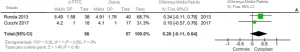
In that same year, Rossini et al. (2015), in turn, carried out a systematic review to verify the effectiveness of orthodontic movements obtained by aesthetic aligners and reported the following findings:
A quantidade média de intrusão relatada foi de 0,72 mm. A extrusão foi o movimento mais difícil de controlar (30% de precisão), seguido pela rotação. A distalização do molar superior revelou a maior previsibilidade (88%) quando foi prescrito um movimento corporal de pelo menos 1,5 mm. Observou-se diminuição do Índice de Little (arco mandibular: 5 mm; arco maxilar: 4 mm) nos arcos de alinhamento.
After that, in 2016, the same authors carried out a systematic review on the accuracy of diagnosis and digital models obtained for orthodontics, with 35 relevant studies, and concluded that so far digital models were “as reliable as traditional plaster models , with high precision, reliability and reproducibility”. In addition, they observed that the identification of the reference points was a limiting factor instead of the measuring device or the software, concluding, however, that digital models could be considered the new gold standard in current practice (ROSSINI et al. , 2016).
Thus, in 2017, Cesur, Omurlu and Ozer (2017) evaluated the accuracy of the digital models produced with the 3D dental scanner compared with a master model and with models obtained from the negative impression of alginate immediately after molding, and of the models obtained at t(0) immediately; t (1) one day; t (2) two days, where the alginate deformation was evaluated separately. Therefore, they made 11 linear measurements of 9 points and found that the arch perimeter was not changed at t (0) = 0hs and t (1) = 24hs, but was changed at t (2) = 48hs and that all models of plaster had a significant difference between the negative scan immediately after the impression. Concluding, therefore, that the measurements of the negative alginate impression, that is, made directly in the mold, have a high degree of precision when compared to those of the positive plaster models or positive impressions.
On the other hand, in 2017, Kamimura et al. (2017) compared silicone molding and scanning in 12 patients, performed by 2 operators with different levels of experience, one with 3 and the other with 16 years of experience, in order to assess the influence of years of experience of professionals in carrying out the molding. For this, they scanned the plaster models on a benchtop scanner. Afterwards, they overlapped the images of the 2 operators and it was observed that the scans had smaller differences compared to the impressions made by the operators, concluding that the digital impression technique is independent of the operator’s experience.
That same year, Lombardo et al. (2017), however, tested the predictability of digital planning with aesthetic aligners in 16 adult patients, totaling 345 teeth analyzed. Thus, through the execution of the setup, the prescribed and actual rotations, the mesiodistal tip and the buccolingual tip were calculated for each tooth and subsequently analyzed by type of tooth. In this aspect, a predictability of 73.6% was finally obtained, reaching the conclusion that “although the tipping movements were effectively achieved, especially in the molars and premolars, the rotation of the lower canines was an extremely unpredictable movement.”
Aslanidou et al. (2017), in turn, reported the case of an adult female patient who had pain in the Temporomandibular Joint (TMJ). Therefore, after performing a Cone Beam Computed Tomography (CBCT), registering the mandibular movements using ultrasound and scanning the arches, a motion tomography was created and, thus, a bite plate was made for the patient already adjusted to your mandibular movements virtually and printed on a 3D printer.
In 2017, Camardella, De Vasconcellos Vilella and Breuning (2017) compared the accuracy of printed models of intraoral scanners, with three different base designs, using two different types of 3D printing: stereolithography and triple jet technology (polyjet). To do so, they overlaid the scan of the printed models with the original file and took measurements on the 3D images. Therefore, they found that models printed by the polyjet method were accurate, regardless of the base format. While the stereolithography prints were accurate with the regular and horseshoe bases with a reinforcing bar, having transverse reduction only with the horseshoe base.
Next year, Becker et al. (2018), in their study, compared the image created of 20 plaster models from eight CBCT and five tabletop scanners available on the market. By superimposing the corresponding images, it was possible to evaluate the accuracy using 6 control points. Data were grouped by scanner and model into box plots, where it was concluded that CBCT systems did not achieve the accuracy of optical scanners, but the accuracy was sufficient for digital planning and forensic purposes (BECKER et al., 2018).
On the other hand, also in 2018, in comparison to a master model, Kim (2018) evaluated the accuracy of models obtained through three printing methods: multijet, colorjet and milling. Therefore, the images were superimposed and 10 measurements were made per group. Finally, it was found that the milling impressions had a difference of 73.05µm±9.64µm, Multijet 84.52µm±4.78µm, and Colorjet 96.05µm±5.43µm, concluding through statistical tests that the milling method obtained greater precision.
That same year, Tepedino et al. (2018) did a retrospective study where 39 adults were treated with a Nuvola® aligner, and digital models were captured at t(0) pre-treatment; t (1) post-treatment and t (S) digital setup for a period of 12 months. In view of this, it was verified that no statistically significant difference was found for all anterior teeth between the predicted movements and the torque achieved. With this, it was concluded that “the system of aesthetic aligners studied was able to produce clinical outcomes comparable to planning the digital configuration in relation to the torque movements of the anterior teeth.”
In the following year, Bocklet et al. (2019) performed an in vitro study where they scanned a fresh human jaw on an industrial tabletop scanner, obtaining a reference STL image with a precision of 3 μm. Then, with 7 intraoral scanners, the authors superimposed the obtained STL images with the reference to compare the accuracy and realism of 4 types of substrate: dentin, resin, amalgam and enamel. With that, they found a range of accuracy and realism that varied between 20μm and 50 μm, concluding that the different substrates interfere with the accuracy of the scans, with the best results presented by dentin, followed by amalgam, resin and enamel. In this context, the authors argued that because enamel has reflective properties, this decreases the accuracy of the scan.
Zhang et al. (2019), in turn, evaluated the influence of printing techniques and layer thickness on the accuracy of the models. So, after scanning a random model on the flatbed scanner, the authors printed models on three types of DLP printers and on an SLA printer, and then scanned them again on a D2000 scanner, overlaying the images for comparison. Therefore, they found results in the range of 50 to 58μm for DLP and 100μm for SLA.
From another perspective, Morris et al. (2019) compared an application-based treatment monitoring system associated with a jugal retractor and a positioner, where the patient filmed his arch to be converted into a 3D model called the DM platform with an intraoral scanner model iTero®. For this, an initial scan was obtained with iTero®, which was sent to Align Technology®, in order to upload it to the DM platform. Then, he performed the treatments on 10 typodonts and with each clear aligner change they scanned and performed the photos and videos with an Iphone 7 and DM accessories. Therefore, the obtained STL images were superimposed for comparison and no significant difference was found between the two scanning techniques.
In this context, Valizadeh et al. (2019) presented a case study of a cloud system that allows the storage and sharing of patient data by several professionals involved with their treatment. Thus, users can convert files such as STL, DICOM and G-code from one format to another so that it is possible to integrate various professionals such as dentists and laboratory technicians, saving time and shortening distances.
Thus, in the following year, Nagy et al. (2020) carried out a study using a fresh maxilla to compare the accuracy of seven scanners and an impression made with polyvinylsiloxane, an impression material of very high stability. However, the authors modified the technique claiming that the risk of failures is minimized with the use of stitches. As the accuracy of the intraoral scanner is commonly evaluated in comparison to the full arch surface, it does not take into account the starting position of the scan. Therefore, the new method takes into account the origin of the scan and calculates the deviation of predefined identical points between references and test models. In this context, the authors concluded that the molding with polyvinylsiloxane was more accurate than the scans due to a deficiency of the scanners in measuring depth, which even occurred in scanners with a confocal microscopy lens that eliminates the blurred area.
Dutton et al. (2020), on the other hand, compared the accuracy of eight intraoral scanners in relation to different substrates. For this, with an industrial scanner and then with the 8 scanners, they scanned a Typodont prepared with different substrates and superimposed the STL images. This done, the authors found that the type of substrate affects the veracity and precision of a scan, where parallel confocal lens scanners were less sensitive to substrate types than triangulation scanners, and that blue light partially nullifies the deviations caused by the bluish translucent enamel substrates.
Therefore, when studying the Digital Light Processing (DLP) printing process, which prints the object inside a photopolymerizable liquid polymer, photopolymerizing polymer layer by layer with a height of 100 μm to 6 μm, from the deviation of light through of a micro mirror system, in 2020, Zhang et al. (2020) found very high resolution printer technologies. Thus, by combining two-photon lithography (LDF and DLP), they created the femtosecond LDF technology (FS-LDF), reaching a layer height of 500 nm. The printing platforms proved to be configurable, with time, light intensity and wavelength parameters being able to be adjusted. Furthermore, with regard to accuracy, the authors argued that it depends on the polymer used, where polyethylene glycol-diacrylate, in addition to being biocompatible, when pure, can be printed in layers of 6μm.
That said, in the same year, Latham et al. (2020) tested the effect of changing the intraoral scanning technique, in order to verify the accuracy of the paths that the operator travels with the scanner to capture images of the surfaces of the teeth and gums. For this purpose, a total of 16 scans were made in a model, with 4 intraoral scanners and 4 different scanning patterns, and later, the images were compared with a master image that was obtained with a very high-quality table scanner accuracy of the same model. With this, the authors found significant differences in the accuracy of the STL images obtained by different scanning patterns, where they concluded that the best path was starting from the 2nd/3rd molar at 45° followed by scanning the lingual to the canine on the opposite side, returning still scanning lingually, but this time at 90°, followed by the occlusal surface and finally the buccal surface, performed at 45° and then at 90°, repeating the process on the opposite posterior sextant. Thus, general comparisons revealed that the approximation of the real between the IOS systems ranged from 46 μm to 119 μm.
Eliasova et al. (2020), in turn, compared four types of 3D model prints: fused deposition modeling (FDM), polyjettechnology (PJ), SLA stereolithography and selective laser sintering (SLS), scanning the models printed by these four technologies with a scanner industrial in order to superimpose them on the original file. Having done this, they found greater roughness in the FDM and PJ models, while the model printed in SLA had a smoother surface, so that the model printed in SLS showed results similar to those of SLA in homogeneity when comparing the perpendicular directions.
So, finally, Lin et al. (2020), in a systematic review covering works from 2015 to 2020, evaluated the clinical effectiveness of the use of clear aligners in Orthodontics and concluded that based on the available evidence, therapy with clear aligners is effective in the management of small malocclusions. However, fixed appliances are more effective in large movements, including better occlusal contacts than clear aligners. However, “aligner treatment is more effective in controlling incisor extrusion than incisor intrusion.”
3. DISCUSSION
In contemporary orthodontics, the scanning of dental arches for metric analysis or the creation of digital setups are routinely performed, as well as 3D printing for the construction of orthodontic and orthopedic appliances that can finally be installed on patients so that their treatment can be carried out.
Therefore, it is noteworthy that intraoral scanners have received some improvements in terms of the precision and accuracy of their scans (ROSSINI et al., 2016; CESUR, OMURLU and OZER, 2017), as well as aligner materials and printing equipment ( CAMARDELLA, DE VASCONCELLOS VILELLA and BREUNING, 2017; LI et al., 2017).
In this context, according to Cesur, Omurlu and Ozer (2017) and Rossini et al. (2016), scanning plus 3D printing proved to be sufficiently accurate in reproducing dental arches compared to models obtained by conventional molding and later obtaining a plaster model, so that Kamimura et al. (2017) found that, regardless of the operator’s experience with impressions, the scans performed with an intraoral scanner obtained files with the same quality standard.
However, Nagy et al. (2020) found a deficiency of scanners in measuring depth and Dutton et al. (2020) observed that the type of substrate affects the veracity and precision of a scan, also identifying that parallel confocal lens scanners and blue light are less sensitive to different types of substrate compared to triangulation scanners.
Furthermore, Latham et al. (2020) observed that the scanning technique also influenced the results when comparing four different standard intraoral scanning routes.
However, Aslanidou et al. (2017) used the technique of superimposing tomography and scanner images, adding mandibular movements to these images on the computer, so that they were able to create and 3D print a pre-adjusted myorelaxing plate for a patient.
That said, regarding clinical studies with the use of aesthetic aligners, Rossini et al. (2015) agreed that the torque movement was well controlled with translation up to 1.5 mm. However, both Rossini et al. (2015) as Lombardo et al. (2017) found difficulties in the rotation movement. Thus, Lombardo et al. (2017) observed that lower canine and premolar rotation had low predictability (LIN et al., 2020 apud ROSSINI, 2015, p. 884).
Thus, on the other hand, Morris et al. (2019) presented a cell phone application that converts the images obtained into an STL file, making it possible to virtually monitor the progress of orthodontic treatment. And Valizadeh et al. (2019) presented a cloud platform that provides agility, organization and registration of patient data exchanges between professionals involved with the respective treatment, eliminating file format barriers and creating manufacturing centers and prosthetic 3D design centers , in order to bring more comfort and speed to patients and professionals involved in the treatment.
4. CONCLUSION
Whereas this article aimed to answer the following guiding question: what technological improvements were identified and developed between May 2015 and December 2020 in the digital flow in orthodontics? The objective was to identify the technological improvements in the digital flow in orthodontics evidenced by the existing literature between the studied period.
In view of this, it was verified that, according to the literature, scanners are more precise and with high accuracy; DLP printers proved to be more efficient and precise with high accuracy; and research with aligners, aesthetic aligner setups and image overlay resources for joint simulation proved to be scarce.
In addition, it was also verified that the use of CCT images isolated and/or superimposed on scanning images is valid for both diagnosis and treatment planning due to the high accuracy and precision; cloud platforms facilitated the exchange of information between professionals related to patient treatment; and cell phone applications that started to produce 3D images of the patient’s arches made it possible to reduce the number of visits to the office.
With this, it can be concluded that with regard to the diagnosis, planning and execution of orthodontic treatments, there have been technological improvements that have increased the accuracy of the images and impressions of the arches, enabled the printing of some devices and made possible the articulation and movements of the jaws in virtual environment, bringing yet another important diagnostic element to the digital workflow in orthodontics.
REFERENCES
ASLANIDOU, Katerina et al. The fabrication of a customized occlusal splint based on the merging of dynamic jaw tracking records, cone beam computed tomography, and CAD-CAM digital impression. Journal of orthodontic science, India, v. 6, n. 3, p. 104-109, jul. 2017. ISSN: 2278-0203. Disponível em: https://www.ncbi.nlm.nih.gov/pmc/articles/PMC5508405/. Acesso em: 20 set. 2020.
BECKER, Kathrin et al. Accuracy and eligibility of CBCT to digitize dental plaster casts. Clinical Oral Investigations, Alemanha, v. 22, n. 4, p. 1817–1823, 2 mai. 2018. ISSN: 1436-3771. Disponível em: http://link.springer.com/10.1007/s00784-017-2277-x. Acesso em: 05 set. 2020.
BOCKLET, Chris et al. Effect of scan substrates on accuracy of 7 intraoral digital impression systems using human maxilla model. Orthod Craniofac Res, Inglaterra, v. 22, Suppl 1, n. S1, p. 168–174, 10 mai. 2019. ISSN: 1601-6343. Disponível em: https://onlinelibrary.wiley.com/doi/abs/10.1111/ocr.12273. Acesso em: 30 set. 2020.
CAMARDELLA, Leonardo Tavares; DE VASCONCELLOS VILELLA, Oswaldo; BREUNING, Hero. Accuracy of printed dental models made with 2 prototype technologies and different designs of model bases. American journal of orthodontics and dentofacial orthopedics, Estados Unidos v. 151, n. 6, p. 1178–1187, 1 jun. 2017. ISSN: 1097-6752. Disponível em: https://dx.doi.org/10.1016/j.ajodo.2017.03.012. Acesso em: 05 set. 2020.
CESUR, Mine Geçgelen; OMURLU, Imran Kurt; OZER, Taha. Evaluation of digital model accuracy and time-dependent deformation of alginate impressions. Nigerian journal of clinical practice, India, v. 20, n. 9, p. 1175–1181, 1 set. 2017. ISSN: 2229-7731. Disponível em: https://doi.org/10.4103/1119-3077.197012. Acesso em: 05 set. 2020.
DE LUCA CANTO, Graziela. et al. Intra-arch dimensional measurement validity of laser-scanned digital dental models compared with the original plaster models: A systematic review. Orthodontics and Craniofacial Research, Inglaterra, v. 18, n. 2, p. 65–76, 1 mai. 2015. ISSN: 1601-6343. Disponível em: https://doi.org/10.1111/ocr.12068. Acesso em: 05 set. 2020.
DUTTON, Ethan et al. The effect different substrates have on the trueness and precision of eight different intraoral scanners. J Esthet Restor Dent, Estados Unidos, v. 32, n. 2, p. 204–218, 1 mar. 2020. ISSN: 1496-4155. Disponível em: https://dx.doi.org/10.1111/jerd.12528. Acesso em: 30 set. 2020.
ELIASOVA, Hana et al. Surface morphology of three-dimensionally printed replicas of upper dental arches. Applied Sciences, Suissa, v. 10, n. 16, 1 ago. 2020. ISSN: 2523-3971. Disponível em: https://doi.org/10.3390/app10165708. Acesso em: 30 set. 2020.
KAMIMURA, Emi et al. In vivo evaluation of inter-operator reproducibility of digital dental and conventional impression techniques. PLoS One, Estados Unidos, v. 12, n. 6, p. e0179188, 21 jun. 2017. ISSN: 1932-6203. Disponível em: https://dx.plos.org/10.1371/journal.pone.0179188. Acesso em: 05 set. 2020.
KIM, Wook-Tae. Accuracy of dental models fabricated by CAD/CAM milling method and 3D printing method. Journal of Oral Research, Chile, v. 7, n. 4, p. 127–133, 27 apr. 2018. ISSN: 0719-2479. Disponível em: https://doi.org/10.17126/joralres.2018.031. Acesso em: 02 set. 2020.
KÜFFER, Maximilian; DRESCHER, Dieter; BECKER, Kathrin. Application of the Digital Workflow in Orofacial Orthopedics and Orthodontics: Printed Appliances with Skeletal Anchorage. Applied Sciences, Suissa, v. 12, n. 8, p. 3820, 10 abr. 2022. ISSN: 2076-3417. Disponível em: https://doi.org/10.3390/app12083820. Acesso em: 07 set. 2020.
LATHAM, Jason et al. Effect of scan pattern on complete-arch scans with 4 digital scanners. The Journal of Prosthetic Dentistry, Estados Unidos, v. 123, n. 1, p. 85–95, jan. 2020. ISSN: 1097-6841. Disponível em: https://dx.doi.org/10.1016/j.prosdent.2019.02.008. Acesso em: 07 set. 2020.
LIN, Chih-Ling et al. Clinical Effectiveness of Using Clear Aligners in Orthodontic Treatment. Taiwanese Journal of Orthodontics, Taiwan, v. 32, n. 3, p. 2, 3 dez. 2020. ISSN: 2708-2636. Disponível em: https://www.tjo.org.tw/tjohttps://www.tjo.org.tw/tjo/vol32/iss3/2. Acesso em: 27 dez. 2020.
LOMBARDO, Luca et al. Predictability of orthodontic movement with orthodontic aligners: a retrospective study. Progress in Orthodontics, Alemanha, v. 18, n. 1, p. 35, 13 dez. 2017. ISSN: 2196-1042. Disponível em: http://progressinorthodontics.springeropen.com/articles/10.1186/s40510-017-0190-0. Acesso em: 06 set. 2020.
MORRIS, Ryan S. et al. Accuracy of Dental Monitoring 3D digital dental models using photograph and video mode. Am J Orthod Dentofacial Orthop, Estados Unidos, v. 156, n. 3, p. 420–428, 2019. ISSN: 1097-6752. Disponível em: https://dx.doi.org/10.1016/j.ajodo.2019.02.014. Acesso em: 07 set. 2020.
NAGY, Zsolt et al. Comparing the trueness of seven intraoral scanners and a physical impression on dentate human maxilla by a novel method. BMC Oral Health, Inglaterra, v. 20, n. 1, p. 97, 7 abr. 2020. ISSN: 1472-6831. Disponível em: https://dx.doi.org/10.1186/s12903-020-01090-x. Acesso em: 30 set. 2020.
REINER, Bruce; SIEGEL, Eliot; CARRINO, John A. Workflow Optimization: Current Trends and Future Directions. Journal of Digital Imaging, Estados Unidos, v. 15, n. 3, p. 141–152, 1 set. 2002. Disponível em: http://link.springer.com/10.1007/s10278-002-0022-7. ISSN: 1618-727X. Acesso em: 27 jul. 2020.
ROSSINI, Gabriele et al. Diagnostic accuracy and measurement sensitivity of digital models for orthodontic purposes: A systematic review. American journal of orthodontics and dentofacial orthopedics, Estados Unidos, v. 149, n. 2, p. 161–70, 1 fev. 2016. Disponível em: https://pubmed.ncbi.nlm.nih.gov/25412265/. ISSN: 1097-6752. Acesso em: 07 set. 2020.
ROSSINI, Gabriele et al. Efficacy of clear aligners in controlling orthodontic tooth movement: A systematic review. Angle Orthodontist, Estados Unidos, v. 85, n. 5, p. 881–889, 1 set. 2015. Disponível em: http://dx.doi.org/10.1016/j.ajodo.2015.06.029. ISSN: 1945-7103. Acesso em: 02 set. 2020.
TEPEDINO, Michele et al. Movement of anterior teeth using clear aligners: a three-dimensional, retrospective evaluation. Progress in Orthodontics, Alemanha, v. 19, n. 1, p. 9, 2 dez. 2018. Disponível em: https://progressinorthodontics.springeropen.com/articles/10.1186/s40510-018-0207-3. ISSN: 2196-1042. Acesso em: 06 set. 2020.
VALIZADEH, Siavash et al. A novel digital dentistry platform based on cloud manufacturing paradigm. International Journal of Computer Integrated Manufacturing, Inglaterra, v. 32, n. 11, p. 1024–1042, 2 nov. 2019. Disponível em: https://www.tandfonline.com/doi/abs/10.1080/0951192X.2019.1686170. ISSN: 1362-3052. Acesso em: 28 abr. 2020.
ZHANG, Jiumeng et al. Digital Light Processing Based Three-dimensional Printing for Medical Applications. International Journal of Bioprinting, Singapura, v. 6, n. 1, p. 12–27, 29 nov. 2020. Disponível em: https://www.ncbi.nlm.nih.gov/pmc/articles/PMC7415858/. ISSN: 2424-8002. Acesso em: 03 dez. 2020.
ZHANG, Zhe-Chen et al. Influence of the three-dimensional printing technique and printing layer thickness on model accuracy. J Orofac Orthop, v. 80, n. 4, p. 194–204, 2019. Disponível em: https://dx.doi.org/10.1007/s00056-019-00180-y. ISSN: 1434-5293. Acesso em: 07 set. 2020.
[1] Specialist in Orthodontics, Specialist in Implantology, Graduated in Dentistry.
[2] Doctor in Orthodontics.
[3] Master in Orthodontics.
[4] Specialist.
[5] Advisor.
Submitted: December, 2021.
Approved: July, 2022.