ORIGINAL ARTICLE
ARAUJO, Priscila Pinto Brandão de [1], NOUER, Paulo Roberto Aranha [2]
ARAUJO, Priscila Pinto Brandão de. NOUER, Paulo Roberto Aranha. Assessment of angular cephalometric measurements for the analysis of vertical growth trend and angle Z in a sample of patients with normal occlusion. Revista Científica Multidisciplinar Núcleo do Conhecimento. Year. 08, Ed. 06, Vol. 01, pp. 44-53. June 2023. ISSN: 2448-0959, Access link: https://www.nucleodoconhecimento.com.br/dentistry/cephalometric-measurements, DOI: 10.32749/nucleodoconhecimento.com.br/dentistry/cephalometric-measurements
ABSTRACT
This study aimed to evaluate the angular measurements and facial characteristics of individuals from the city of Rio de Janeiro, Brazil, using cephalometry. For this study, 531 male individuals from military organizations in Rio de Janeiro were submitted to the clinical record of the Master’s degree in Orthodontics course of the Research Center – São Leopoldo Mandic. Of this sample, it was observed that 22 individuals had normal occlusion and had never undergone orthodontic treatment. In these patients, lateral cephalograms were taken and the cephalometric measurements were evaluated: Y-axis, NS.Go-Me, SN.PLO, FMA, SN-Go-Gn, angle Z, among others. The data obtained were subjected to analysis with a significance level of 5% (p<0.05) and descriptive statistics and the Dahlberg error were used. The results showed that individuals with normal occlusion from the city of Rio de Janeiro presented these cephalometric characteristics within the normal standards as already described in the literature.
Keywords: Vertical growth, Orthodontics, Cephalometry, Prevalence.
INTRODUCTION
Orthodontists have always been interested in knowing the characteristics of the population to which they offer their services, since some malocclusions are more frequent in certain racial groups. Therefore, studies should be carried out to individualize the characteristics of certain populations so that the diagnosis and treatment plan for orthodontic patients follows the facial patterns of that particular population (REIS, 2006). In this way, the individualization of cephalometric norms for different races is of extreme importance (FERNANDES, 2017).
Angle (1899) classified malocclusions into class I, II, or III based on the position of the permanent first molar in relation to the permanent first molar, classifying individuals as class I when the mesio-vestibular cusp of the upper first molar occludes in the mesio-vestibular groove of the lower first molar, class II individuals when the mesio-vestibular cusp of the upper first molar occludes in front of the mesio-vestibular groove of the lower first molar, and class III individuals when the mesio-vestibular cusp of the upper first molar occludes behind the mesio-vestibular groove of the lower first molar.
The emergence of lateral cephalograms, with Broadbent (1931), made it possible to assess the various cephalometric measurements of interest to orthodontists, encouraging numerous professionals and institutions to develop techniques and systematics for characterizing the skeletal architecture of the face. From the grouping of the various cephalometric measurements, cephalometric analyses emerged, which provide information on the sizes and shapes of the craniofacial components, their relative positions and orientations.
Downs (1948) determined standards of normality for the skull and face through cephalometric analysis, obtained from 20 young people with normal occlusion between 12 and 17 years of both genders, observing that when the “Y” axis of growth deviated from the average obtained, it indicated a horizontal or vertical growth, according to the angle found, concluding that the results obtained varied greatly from the measurements studied, and therefore, the importance of evaluating the skeletal pattern individually.
Tweed (1946) described the angles IMPA, FMA, and FMIA, determining that the calculation of the discrepancy of his cephalometric analysis should be obtained from the IMPA and that the normal values for FMA and FMIA are respectively 25º and 65º, with the IMPA being 90º; when the FMA increased or decreased, there should be compensation in the value of the IMPA and would demonstrate the vertical growth of the patient. He thus determined that each individual should be evaluated individually, observing the position of the lower incisor in the basal bone.
Merrifield (1966), studying 120 lateral cephalometric radiographs, established a tangent line to the soft chin and the most prominent lip, upper or lower, until it reached the Frankfurt horizontal plane, forming the Z angle. He obtained an average value of 80º for adults and 78º for individuals between 11 and 15 years old.
Based on radiographic cephalometry, several cephalometric analyses have been developed and obtained from various ethnic groups. However, such analyses when applied to Brazilian individuals from different Brazilian regions may present comparison errors. With this, we should seek values that characterize the differentiation of each ethnic and regional group, in order to use correct diagnostic and treatment plan standards. Based on what was exposed, this study aimed to evaluate malocclusion in a population of individuals from the Municipality of Rio de Janeiro as well as to individualize cephalometric patterns of individuals with normal occlusion.
MATERIAL AND METHODS:
The examination of the soldiers was carried out only after obtaining the approval of the Ethics Committee in Research of the Centro de Estudos Odontológicos – São Leopoldo Mandic (Protocol 05/322) and the authorization of those selected with clinically normal occlusion and only then was it started.
MATERIAL
The sample for this study was obtained from the population of soldiers belonging to the military organizations of the state of Rio de Janeiro – RJ. 531 individuals aged 18 to 25 years, male, feodermas, born in the municipality of Rio de Janeiro and not submitted to any type of orthodontic treatment were evaluated.
From the sample of 531 soldiers, those who had an occlusion considered clinically normal were selected, following the following inclusion criteria:
- Adequate oral health, without cavities, periodontal problems, or pathologies;
- Passive lip seal;
- Vertical and horizontal symmetry;
- Absence of harmful oral habits;
- Coinciding midline;
- Absence of crowding and dental rotations in all segments;
- Presence of all permanent teeth, except for third molars;
- Normal occlusion key molar relationship;
- Class I canine relationship;
- Absence of noises/ clicks in the TMJ;
- Incisor and canine guidance without interferences.
Of the total, 49 individuals were selected who had these characteristics, however only 22 individuals agreed to participate in the research.
OBTAINING TELERRADIOGRAPHS
After selecting the 22 individuals with clinically normal occlusion, they were referred to a dental documentation center (COR, Rio de Janeiro, Brazil) for head radiographs. Using the technique recommended by Broadbent (1931), of cephalostat positioning.
CEPHALOMETRIC TRACING
On each teleradiography, a sheet of transparent acetate paper “ultraphan” (GAC, Bohemia, New York, USA) of size 17.5 x 17.5 cm, thickness 0.07 mm was adapted and then with a 0.7 mm pencil (Faber-Castell, São Carlos, Brazil) and a “template” (GAC, Bohemia, New York, USA) the cephalogram was traced on a negative viewer (VH, Araraquara, São Paulo, Brazil) in a dark room to better highlight the anatomical structures. The tracings were performed by a single researcher.
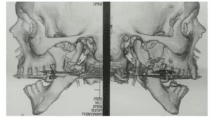
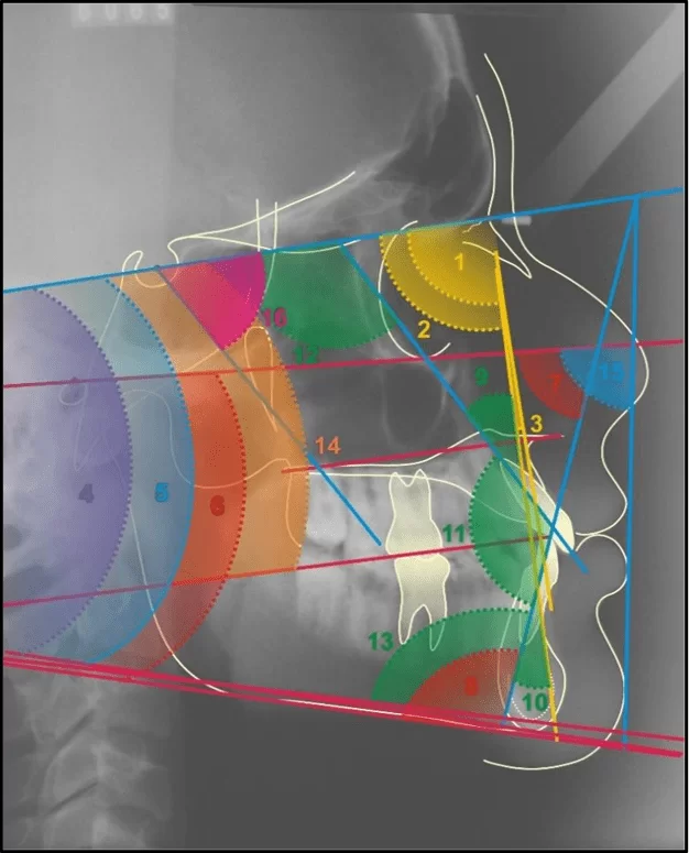
The cephalograms were traced, delimiting the anatomical structures of the skull and face, where lines and planes were drawn which gave rise to the angular cephalometric measurements (figures 1), which are part of the protocol of the Master’s Degree in Orthodontics course of the CPO – São Leopoldo Mandic according to the Cephalometry Manual recommended by Nouer (NOUER,2003).
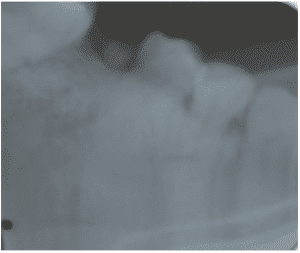
Figure 1. Obtaining angular cephalometric measurements
STATISTICAL ANALYSIS
In the statistical analysis, the significance level was set at 5% (p<0.05), and descriptive statistics (minimum, mean, maximum, and standard deviation) and the Dahlberg error were utilized.
RESULTS
PREVALENCE OF MALOCCLUSION
The results of the prevalence of malocclusion are presented in Table 1. The sample was divided according to the malocclusion classification, resulting in a higher prevalence of Class I malocclusion at 44.3%, followed by Class II (29.5%), where Class II, division 1, prevailed, Class III (17.2%), and normal occlusion (9.2%).
Table 1. Distribution of patient classification based on malocclusion
| Classification | Normal | Class I | Class II | Class III | Total |
| 0(cl. I) | 49 (100) | 235 (100) | – | – | 284 (53,5) |
| 1(cl. II d1) | – | – | 64 (41,0) | – | 64 (12,1) |
| 2(cl. II d2) | – | – | 26 (16,7) | – | 26 (4,9) |
| 3(cl. III ) | – | – | – | 63 (69,2) | 63 (11,9) |
| 4(cl.II d1 sub.D) | – | – | 25 (16,0) | – | 25 (4,7) |
| 5(cl. II d1 sub.E) | – | – | 26 (16,7) | – | 26 (4,9) |
| 6(cl. II d2 sub.D) | – | – | 12 (7,7) | – | 12 (2,3) |
| 7(cl. II d2 sub.E) | – | – | 3 (1,9) | – | 3 (0,6) |
| 8(cl. III sub. D) | – | – | – | 16 (17,6) | 16 (3,0) |
| 9(cl. III sub.E) | – | – | – | 12 (13,2) | 12 (2,3) |
| Total | 49 (100) | 235 (100) | 156 (100) | 91 (100) | 531 (100) |
Source: Prepared by the authors, 2005.
CEPHALOMETRIC ANGULAR MEASURES ANALYZED FOR ASSESSING VERTICAL GROWTH TENDENCY IN PATIENTS WITH NORMAL OCCLUSION AND ANGLE Z
Among the analyzed angular measures, it was observed that the mean values for the entire sample fall within a pattern of vertical growth normalcy for individuals classified with a normal occlusion.
Table 2. Mean, standard deviation, minimum, maximum, and median of all vertical angular measures
| n | mean | st.dev. | min | median | max | |
| angle“Y” (º) | 22 | 67,0 | 5,0 | 57 | 67,5 | 80 |
| NS.Go-Me (º) | 22 | 29,7 | 5,6 | 20 | 32 | 40 |
| FMA (º) | 22 | 22,8 | 5,4 | 15 | 22,5 | 32 |
| FMIA (º) | 22 | 57,0 | 5,7 | 47 | 56,5 | 70 |
| SN.GO-GN (º) | 22 | 28,4 | 5,7 | 18 | 30 | 38 |
| SN.PLO (º) | 22 | 11,3 | 3,7 | 4 | 11 | 20 |
| angle Z (º) | 22 | 73,8 | 6,9 | 60 | 72 | 85 |
Source: Prepared by the authors, 2005. Note: No significant difference (p>0.05).
DISCUSSION
Functional and aesthetic results can be achieved harmoniously when the professional is capable of identifying functional and skeletal disharmonies that affect bone growth and determine the patient’s occlusion. It is crucial to understand what is considered normal for a specific population group. Orthodontic-orthopedic techniques can be used more efficiently, considering the individual characteristics of each patient (TREVISAN, 2006).
The cephalometric angular measures indicating the individual’s vertical growth tendency evaluated in this study included the “Y” angle², Ns. Go-Me, FMA, Sn.Go-Gn, and SN.Plo. Our found values were: “Y” angle = 67º, Ns.Go-Me = 29.7º, FMA = 22.8º, SN.Go.Gn = 28.4º, SN.PLO = 11.3º (Table 2). These values indicate a horizontal growth tendency in patients, average values suggesting growth within the normal standards of the studied sample.
We observed that the variable Z angle did not show a significant result when evaluated (Tables 1). The value established by the Tweed Foundation was determined by Merrifield (78º). The average value for the sample in group 1, with normal occlusion, was 73.8º ±6.9º (MERRIFIELD, 1966).
The analysis of angular measures assessing vertical growth tendency combined with the evaluation of the Z angle showed for this sample that in patients with a normal occlusion, a pattern of horizontal growth and a profile within normality is most commonly observed. This is consistent with various works in the literature (SILVA, 2004).
CONCLUSION
We understand that several authors have found various cephalometric measures based on the study of specific populations, which may not always reflect the characteristics of all individuals worldwide or within diverse Brazilian populations. Hence, it is essential to individually study cephalometric measures in various populations, both within Brazil and worldwide, so that each population with its specific characteristics receives a correct diagnosis and an individualized orthodontic treatment plan.
REFERENCES
ANGLE, E.H. Classification of malocclusion. Dental Cosmos, v. 41, pp. 248-264, 1899. Disponível em: <https://quod.lib.umich.edu/d/dencos/acf8385.0041.001/267:56?page=root;size=100;view=pdf>. Acesso em: 07 jun. 2023.
BROADBENT, BH. et al. A new X-ray technique and its application to orthodontia. The Angle Orthod, v.1, n. 2, pp. 45-66, 1931. Disponível em: <https://www.scienceopen.com/document?vid=8c188ea3-8833-41ee-9b68-1384f1c706cb>. Acesso em: 07 jun. 2023.
DOWNS, WB. Variations in facial relationships: their significance in treatment and prognosis. American Journal of Orthodontics, v. 34(10), pp. 812-840, 1948. Disponível em: <https://pubmed.ncbi.nlm.nih.gov/18882558/>. Acesso em: 07 jun. 2023.
FERNANDES, L.P.L. Alterações das variáveis cefalométricas nos diferentes grupos étnicos [dissertação]. Coimbra: Universidade de Coimbra, 2017. Disponível em: <https://estudogeral.uc.pt/handle/10316/82760>. Acesso em: 07 jun. 2023.
MERRIFIELD, LL. The profile line as an aid in critically evaluating facial estetics. Am. J. Orthod, Saint Louis, v. 52, n. 11, p. 804-822, 1966. Disponível em: <https://www.sciencedirect.com/science/article/abs/pii/0002941666902508?via%3Dihub>. Acesso em: 07 jun. 2023.
NOUER, P.R.A. Cefalometria aplicada em radiologia e ortodontia. São Paulo: Santos; 2003.
REIS, S.A.B. et al. Análise facial subjetiva. Dental Press Journal of Orthodontics, Maringá, v. 11, n. 5, pp. 159-172, 2006. Disponível em: <https://www.scielo.br/j/dpress/a/vcNJFKLcHG8ZsCh747q9Hxy/?format=pdf&lang=pt>. Acesso em: 07 jun. 2023.
SILVA, OP. et al. Padrão cefalométrico de brasileiros leucodermas portadores de oclusão “normal”. Rev. Dental Press Ort. Ortop Facial, v. 9, n. 1, pp. 59-78, 2004.
TREVISAN, F.; GIL, C.T.L.A. Análise fotogramétrica e subjetiva do perfil facial de indivíduos com oclusão normal. Dental Press Journal of Orthodontics, v. 11, n. 4, pp. 24-35, 2006, Disponível em: <https://www.scielo.br/j/dpress/a/5xnXRw7P8YxmHz4QxyRDnQw/?lang=pt>. Acesso em: 07 jun. 2023.
TWEED, CH. The Frankfort-mandibular plane angle in orthodontic diagnosis, classification, treatment planning, and prognosis. Am J Orthod Oral Surg, v. 32, n. 4, pp. 175-230, 1946. Disponível em: <10.1016/0096-6347(46)90001-4>. Acesso em: 07 jun. 2023.
[1] Advanced Studies in Orthodontics and Functional Maxillary Orthopedics, Specialization in Public Health, Master’s in Orthodontics, Ph.D. in Orthodontics. ORCID: 0000-0002-5514-0911. Currículo Lattes: http://lattes.cnpq.br/1678395879499706.
[2] Advisor. Specialization in Orthodontics, Specialization in Radiology, Ph.D. in Dentistry, and Postdoctoral Studies in Orthodontics. ORCID: 0000-0003-3239-4780. Currículo Lattes: http://lattes.cnpq.br/3913946805965518.
Submitted: March 21, 2023.
Approved: May 31, 2023.