ORIGINAL ARTICLE
ARAGÃO, José Aderval [1], MATOS, Ícaro Quintela [2], CARVALHO, Higor Dantas Gonçalves [3], ARAGÃO, Iapunira Catarina Sant’Anna [4], ARAGÃO, Felipe Matheus Sant’Anna [5], LOURENÇO, Bárbara Costa [6], FEITOSA, Vera Lúcia Correa [7], REIS, Francisco Prado [8]
ARAGÃO, José Aderval, et al. Sexual dimorphism based on the sternum bone morphometry in human fetuses: a pilot study. Revista Científica Multidisciplinar Núcleo do Conhecimento. Year. 08, Ed. 04, Vol. 01, pp. 115-128. April 2023. ISSN: 2448-0959, Access link: https://www.nucleodoconhecimento.com.br/health/sexual-dimorphism, DOI: 10.32749/nucleodoconhecimento.com.br/health/sexual-dimorphism
ABSTRACT
The recognition and determination of sex from fragments and remnants of human skeletons has been, until now, a complex task for forensic experts and anthropologists. For the former, it becomes crucial in cases of mass disasters, catastrophes, such as terrorist acts and wars. The study with bones of the pelvis, skull, femur and other long bones has significantly contributed to analyze the determination of sex in these situations. The study of sternum dimorphism can become one more element that may collaborate with this type of knowledge. The present study sought to investigate, in human fetuses, the sexual dimorphism of the sternum, based on its perimeter measurements, which were treated using statistical methods. For this purpose, three different observers used a digital pachymeter with a precision of 0.01mm and the average of the measurements obtained was calculated. Differences in average were evaluated using the t-test and Mann-Whitney test, as well as discriminant analysis. Findings from measurements of the sternum in human fetuses did not seem to us to be a reliable indicator of sex determination. We hope that further studies with a larger number of samples and the use of other possible techniques will also collaborate with the subject.
Keywords: Skeleton sex determination, Sternum, Anthropometry, Biometric Identification, Discriminant Analysis, Forensic Medicine.
INTRODUCTION
Variations in sternum size are common among individuals (GOODMAN et al., 1983). Morphometric data regarding the sternum has become important in forensic medicine, in particular, for identifying the sex of people victims of mass disasters, in whom the traces of human skeletons are precarious (SINGH et al., 2012; SINGH and PATHAK, 2013). The comparison between the sexes of the relationship between the length of the sternum and the manubrium was pioneered in forensic radiology by Wenzel (1788) and was followed by Ashley (1956). Dwight (1881) and Hyrtl (1893) recognized and reported a 1:2 ratio pattern in women and 2:1 in men. This led to the creation of Hyrtl’s Law, stating that: the length of the female manubrium exceeds half the length of the mesosternum and the body length of the male sternum is at least twice the length of the manubrium (DWIGHT, 1890). Hunnargi et al. (2009), measuring 115 sternums of the Maharashtrian population of India, tested the viability of Hyrtl’s Law and reported that only 18.7% of the sample corresponded to the established by this Law. Ramadan et al. (2010), using computed tomography of the thorax and comparing their findings to that described by Hyrtl’s Law, found an accuracy of 86% for women and 34% for men.
Studies with the aim of identifying the sex of individuals based on the length and width of the sternum were carried out by authors such as Gautam et al. (2003), Atal et al. (2009), Osunwoke et al. (2010), Sing et al. (2012); Pathak and Singh (2012); Kaneriya et al. (2013); Singh and Pathak (2013) and Changani et al. (2014). These authors admitted the impossibility of determining a value that distinguished the sexes and considered their findings as unsatisfactory and of low efficacy. Recently, more elaborate mathematical methods such as logistic regressions and discriminant analysis have been used (MACALUSO et al., 2010; PUTTABANTHI et al., 2012). Darwish et al. (2017) studying 60 CT scans of the chest of adult Egyptians, performed a multiple regression analysis with the length of the manubrium and body of the sternum; width of the manubrium, third and fourth sternebrae and obtained an accuracy of 96.67%.
Mukhopadhyay (2010) carried out a discriminant analysis of the measurements of the width of the jugular notch, from the sternum to the level of the 4th rib and the length of the posterior curvature, the author highlighted the 100% accuracy in the diagnosis of sex. Several studies using different mathematical treatments have found sexual dimorphism of the sternum in different regions, such as: South India (CHANDRAKANTH; KANCHAN; KRISHAN, 2014), South Africa (MACALUSO et al., 2010), Bengal (MUKHOPADHYAY, 2010), Northwest Bulgaria (TONEVA; NIKOLOVA, 2014).
Similar studies have not been common in the Brazilian population (REBELO et al., 2014; SILVA et al., 2021). Thus, there is a timely need for studies on the sexual dimorphism of the sternum in the Brazilian population, considering the value of the measurements of the sternum demonstrated in the most different populations (ROSS; UBELAKER; KIMMERLE, 2011). The recurrent use of literature on adults is because studies on the subject in fetuses are infrequent. Aragão et al. (2021), carried out a study on the sternal index in human fetuses as an indicator of sexual dimorphism.
MATERIAL AND METHOD
Thirty sternums, 15 from male and 15 female human fetuses, were dissected, removed, cleaned and the costosternal articulations preserved. The fetuses were aged 20.9 to 36.8 weeks, a mean age of 27.63±4.5 and median of 27.1 weeks. The thirty fetuses in the study were available at the Anatomy Laboratory, chosen at random. These fetuses were obtained in accordance with Law No. 8501 of November 30, 1992, which addresses the use of unclaimed cadavers for use in research studies. The Research Ethics Committee of the Federal University of Sergipe opinion number 1.462.381 approved these studies.
In the selection, only fetuses with complete rib cages were considered, while those who showed any kind of alteration in their spine, including kyphosis, lordosis, scoliosis, or any visible malformation of the anterior thoracic area, were excluded.
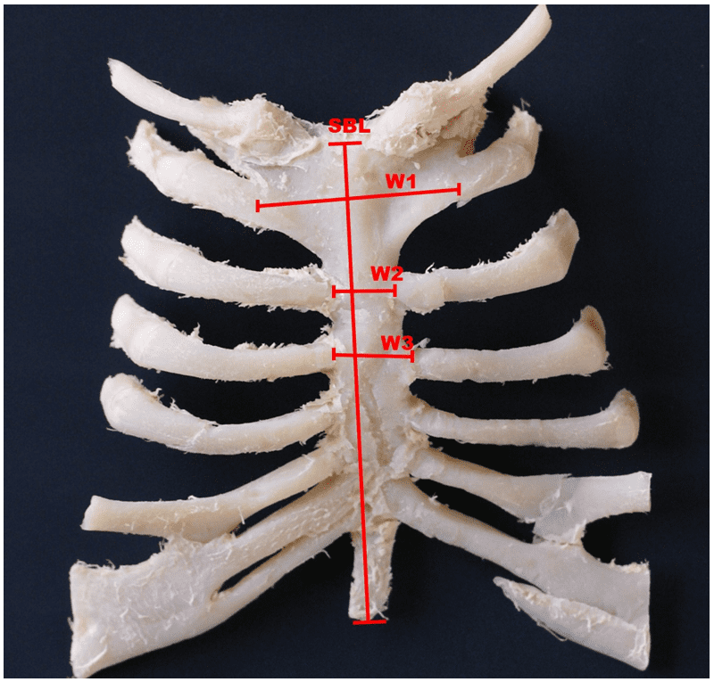
Three independent observers performed the measurements with the aid of a digital caliper with a precision of 0.01 mm and measurements between each of them were averaged. There was no recordable discrepancy level. Sternal Bone Length (SBL) (distance between the jugular notch on the manubrium of the sternum to the apex of the cartilaginous xiphoid process). Sternal widths were measured from the notch of the costosternal joints: 1 (W1) (distance between the costal notches of the first ribs); width 2 (W2) (distance between the costal notches of the second ribs); and 3 (W3) (distance between the costal notches of the third ribs) (Figure 1).
Figure 1. Morphometric measurements of the sternum
Data analyses were described by mean, standard deviation, minimum and maximum. Mean differences were evaluated using the Welch’s unpaired t-test and the Mann-Whitney test. Welch’s unpaired t-test was used because its assumptions of normality (Shapiro-Wilks Test) and equality of variances (Levene Test) were met. Under those conditions, Welch’s T test is more powerful than Mann-Whitney test (GERKE and RANDLES, 2010). To assess the discriminant function, all models including sternum length, width 1, width 2, and width 3 were tested, such as individual models, pairwise models, three-way models, and comprehensive models. For each model, we computed non-standardized coefficients, standardized coefficients, and centroids based on gender. Discriminant Analysis has univariate and multivariate normality, equality of variances, and a non-singular scatter matrix as assumptions, which were met in all models (SHARMA and PALIWAL, 2013). The software used was the R Core Team 2018 and the significance level adopted was 5%.
RESULTS
The descriptive statistics of the four variables (sternum length, width 1, width 2 and width 3) are presented in Table 1. The results showed that all male variables had slightly higher means than female ones, but without statistically significant difference between genders for any of the variables, according to the t-test or the Mann-Whitney test (p>0.05). Analyzing the standardized coefficients, we can infer from the model with all the variables that the parameters that most influenced sexual differentiation were sternum width 3 (1.847), sternum width 2 (-0.905), sternum width 1 (-0.657) and length of the sternum (0.024).
Table 1. Descriptive statistics of the sternum measurements
| Female
(n=15) |
Male
(n=15) |
|||||
| Mean (SD) | Med [Min-Max] | Mean (SD) | Med [Min-Max] | t (p-value) | p-value* | |
| Sternumlength | 52.28 (7.98) | 52.66 [37.31-63.02] | 53.23 (12.81) | 54.58 [35.27-76.61] | 0.24 (0.809) | 0.885 |
| Sternumwidth 1 | 13.36 (3.09) | 12 [10.34-19.13] | 13.48 (2.48) | 13.28 [9.98-18.18] | 0.12 (0.907) | 0.917 |
| Sternumwidth 2 | 11.84 (2.91) | 11.47 [8.15-16.76] | 11.96 (2.35) | 11.93 [7.39-15.48] | 0.12 (0.902) | 0.756 |
| Sternumwidth 3 | 10.16 (2.19) | 9.70 [6.83-13.58] | 10.55 (1.85) | 10.68 [7.32-13.85] | 0.53 (0.599) | 0.520 |
Test t; * Mann-Whitney test; SD – Standard Deviation; Med – Median.
Source: Authors.
The centroids of each sex showed a greater distance in the models with widths 1, 2, and 3 from the sternum and in the model with all variables (0.146). Consequently, the standardized and non-standardized coefficients for each variable, as well as constants for all possible functions created by combining the four variables, were calculated with the segment point set at 0. As such, when the discriminant score function result was higher than 0, it was male, while a value lower than 0 was considered female (Table 2).
Table 2. Coefficients of the discriminant function analysis with the limit points
| Coefficient
(non-standardized) |
Coefficient
(standardized) |
Centroid | ||
| F | M | |||
| Variable | ||||
| Sternumlength | 0.094 | 1.000 | -0.045 | 0.045 |
| Constant | -4.942 | |||
| Sternumwidth 1 | 0.357 | 1.000 | -0.021 | 0.021 |
| Constant | -4.791 | |||
| Sternumwidth 2 | 0.379 | 1.000 | -0.023 | 0.023 |
| Constant | -4.507 | |||
| Sternumwidth 3 | 0.494 | 1.000 | -0.097 | 0.097 |
| Constant | -5.120 | |||
| Sternumlength | 0.118 | 1.258 | -0.047 | 0.047 |
| Sternumwidth 1 | -0.154 | -0.431 | ||
| Constant | -4.149 | |||
| Sternumlength | 0.117 | 1.247 | -0.046 | 0.046 |
| Sternumwidth 2 | -0.153 | -0.403 | ||
| Constant | -4.348 | |||
| Sternumlength | -0.058 | -0.624 | -0.107 | 0.107 |
| Sternumwidth 3 | 0.683 | 1.382 | ||
| Constant | -3.993 | |||
| Sternumwidth 1 | 0.173 | 0.484 | -0.024 | 0.024 |
| Sternumwidth 2 | 0.235 | 0.620 | ||
| Constant | -5.112 | |||
| Sternumwidth 1 | -0.321 | -0.899 | -0.122 | 0.122 |
| Sternumwidth 3 | 0.720 | 1.457 | ||
| Constant | -3.153 | |||
| Sternumwidth 2 | -0.416 | -1.100 | -0.131 | 0.131 |
| Sternumwidth 3 | 0.795 | 1.609 | ||
| Constant | -3.280 | |||
| Sternumlength | 0.130 | 1.390 | -0.048 | 0.048 |
| Sternumwidth 1 | -0.123 | -0.344 | ||
| Sternumwidth 2 | -0.114 | -0.300 | ||
| Constant | -3.871 | |||
| Sternumlength | -0.024 | -0.254 | -0.124 | 0.124 |
| Sternumwidth 1 | -0.287 | -0.803 | ||
| Sternumwidth 3 | 0.775 | 1.568 | ||
| Constant | -2.924 | |||
| Sternumlength | -0.021 | -0.222 | -0.133 | 0.133 |
| Sternumwidth 2 | -0.386 | -1.019 | ||
| Sternumwidth 3 | 0.843 | 1.705 | ||
| Constant | -3.040 | |||
| Sternumwidth 1 | -0.232 | -0.649 | -0.146 | 0.146 |
| Sternumwidth 2 | -0.340 | -0.899 | ||
| Sternumwidth 3 | 0.917 | 1.855 | ||
| Constant | -2.335 | |||
| Sternumlength | 0.002 | 0.024 | -0.146 | 0.146 |
| Sternumwidth 1 | -0.234 | -0.657 | ||
| Sternumwidth 2 | -0.343 | -0.905 | ||
| Sternumwidth 3 | 0.913 | 1.847 | ||
| Constant | -2.350 | |||
Source: Authors.
The accuracy of the Discriminant Functions (DF) was tested with the obtained functions (Table 3). The highest level of accuracy for females was observed in several models (accuracy=60%), however there was not much variation since the other models reached an accuracy of 53%. It was possible to infer a slight predictive homogeneity for the female gender. The best results (60%) were observed at widths 1 and 3 of the examined sternums.
Table 3. Accuracy of the discriminant functions for sex determination
| F | M | Total | |
| SBL | 53.3% | 53.3% | 53.3% |
| W1 | 60.0% | 40.0% | 50.0% |
| W2 | 53.3% | 53.3% | 53.3% |
| W3 | 60.0% | 60.0% | 60.0% |
| SBL, W1 | 53.3% | 46.7% | 50.0% |
| SBL, W2 | 60.0% | 53.3% | 56.7% |
| SBL, W3 | 60.0% | 60.0% | 60.0% |
| W1, W2 | 53.3% | 53.3% | 53.3% |
| W1, W3 | 60.0% | 53.3% | 56.7% |
| W2, W3 | 60.0% | 73.3% | 66.7% |
| SBL, W1, W2 | 53.3% | 46.7% | 50.0% |
| SBL, W1, W3 | 60.0% | 53.3% | 56.7% |
| SBL, W2, W3 | 60.0% | 73.3% | 66.7% |
| W1, W2, W3 | 60.0% | 66.7% | 63.3% |
| SBL,W1, W2,W3 | 60.0% | 66.7% | 63.3% |
Source: Authors.
Results for men were more dispersed than for women, with an accuracy of 40% to 73.3%. The most accurate models included sternal widths 1 and 2, as well as sternal length and sternal widths 2 and 3, which achieved 73.3% accuracy. In single models, sternal width 3 had the highest accuracy (60%). The highest total accuracy was obtained using variables W2, W3 or SBL, W2 and W3, both functions obtained 66.7% of correct sex diagnosis. When making a function with only one variable, W3 achieved the highest accuracy with 60% accuracy. The isolated variable with the lowest accuracy was W1 with only 50% accuracy. According to the discriminant function, determined through the equations, DF = – 0.021*SBL – 0.386*L2 + 0.843*L3 – 3.040 and DF = – 0.416*W2 + 0.795*W3 – 3.280, respectively, was a low level of accuracy to determine the sex of fetuses by measurements of the sternum.
DISCUSSION
The present study evaluated four measurements of the sternum in fetuses, with the aim of establishing a relationship between bone morphometry and sex. Our findings showed that it was not possible to develop an effective for sex determination based on sternum morphometry of human fetuses.
Regarding the Mann-Whitney test, in the present study, it had different results compared to similar studies (CHANDRAKANTH; KANCHAN; KRISHAN, 2014; TONEVA; NIKOLOVA, 2014; DARWISH et al., 2017). These authors found a statistically significant difference between genders. It is possible that this occurred because the fetuses have a low sexual dimorphism in their measurements, which may make it difficult to accurately determine the sex with only the sternum measurements.
The values obtained for the discriminant functions, developed in the current study, using SBL, W2 and W3 or W2 and W3, had an accuracy of 73.3% for men and 60% for women. These values are low, when compared with the value reported by Singh et al. (2012), who obtained an accuracy of 84%, studying 343 sternums of cadavers from North India. Macaluso; Lucena (2014), in Spain, in a sample of 116 sternums, developed a discriminant function with 89.7% accuracy. We admit that differences in accuracy may possibly be related to the use of fetuses.
Despite this, the accuracy values of the present study were comparatively superior to those that used simpler mathematical methods, such as the demarcation of limit points. Changani et al. (2014) used the demarcation of limit points for the length of the sternum and obtained 43.86% accuracy, with most sample values in the overlapping zone. Ramadan et al. (2010) using the length of the manubrium were able to correctly determine 69% and 60% of the female and male sternums, respectively. The low efficiency of these studies may have occurred due to most of the population being in an overlapping zone, which may have made it difficult to demarcate border points, as demonstrated by Atal; Murari; Naik (2009).
CONCLUSIONS
In this study, an accuracy of 73.3% for males and 60% for females was obtained using the variables W2, W3 or SBL, W2, W3. Based on this, we can conclude that the measurements of the sternum of fetuses did not provide us with definitive values regarding the sex of the individual. We hope that more studies with a greater number of samples and the use of other possible techniques will also contribute to this subject.
REFERENCES
ARAGÃO, J. A.; GONÇALVES CARVALHO, H. D.; MATOS, I. Q.; CAVALCANTI, R. S.; SANT’ANNA ARAGÃO, I. C.; SANT’ANNA ARAGÃO, F. M.; MARASSI, P. H. A.; CARDOSO, P.; LOURENÇO, B. C.; REIS, F. P. Sternal index in human fetuses as an indicator of sexual dimorphism. J Morphol Sci. n. 38, p. 321-324, 2021.
ASHLEY, G. T. A comparison of human and anthropoid mesosterna. Am J Phys Anthropol., vol. 14, n. 3, p. 449-65, 1956.
ATAL, D. K.; MURARI, A.; NAIK, S. K. Gender differentiation from sternal widths. J Ind Acad Forensic Med., vol. 30, n. 4, p. 198-201, 2009.
CHANDRAKANTH, H. V.; KANCHAN, T.; KRISHAN, K. Osteometric analysis for sexing of modern sternum – An autopsy study from South India. Leg Med (Tokyo), vol. 16, n. 6, p. 350-6, 2014.
CHANGANI, M. V.; JAVIA, M. D.; VARMA, K. A. Determination of sex from various measurements of human sternum & manubrium in Gujarat population. J Red Med Den Sci, vol. 2, n. 1, p. 59-65, 2014.
DARWISH, R. T.; ABDEL-AZIZ, M. H. E. L.; NEKIEDY, A. M.; SOBHZK. Sex determination from chest measurements in a sample of Egyptian adults using Multislice computed tomography. J Forensic Leg Med, vol. 52, p. 154-8, 2017.
DWIGHT, T. Sternum as an Index of Sex, Height, and Age. J AnatPhysiol., vol. 24, n. Pt 4, p. 527-35, 1890.
DWIGHT, T. The Sternum as an Index of Sex and Age. J AnatPhysiol., vol. 15, n. Pt 3, p. 327-30, 1881.
GAUTAM, R. S.; SHAH, G. V.; JADAV, H. R.; GOHIL, B. J. The human sternum: as an index of age and sex. J Anat Soc Ind., vol. 52, n. 1, p. 20-3, 2003.
GERKE, T. A.; RANDLES, R. H. A method for resolving ties in asymptotic relative efficiency. Statistics & probability letters, vol. 1, n. 80, p. 1065-9, 2010.
GOODMAN, L. R.; TEPLICK, S. K.; KAY, H. Computed tomography of the normal sternum. AJR Am J Roentgenol, vol. 141, n. 2, p. 219-23, 1983.
HUNNARGI, S. A.; MENEZES, R. G.; KANCHAN, T.; LOBO, S. W., BINU, V. S.; UYSAL, S.; KUMAR, H. R.; BARAL, P.; HEREKAR, N. G.; GARG, R. K. Sexual dimorphism of the human sternum in a Maharashtrian population of India: a morphometric analysis. Leg Med (Tokyo), vol. 10, n. 1, p. 6-10, 2009.
HYRTL, J. Handbuch der topographischen anatomic percentage. Vienna: Wilhelm Braumuller, 1893.
KANERIYA, D.; SUTHAR, K.; PATEL, V.; UMARVANSHI, B.; MEHTA, C.; TAILOR, C. Morphometric study of sternum for determination of sex. Cibtech J Bio-Protocols, vol. 2, n. 2, p. 6-13, 2013.
MACALUSO, P. J.; LUCENA, J. Estimation of sex from sternal dimensions derived from chest plate radiographs in contemporary Spaniards. Int J Legal Med., vol. 128, n. 2, p. 389-95, 1893.
MACALUSO, P. J. Et al. The efficacy of sternal measurements for sex estimation in South African blacks. Forensic Sci Int., vol. 202, n. 1-3, p. 111.e1-7, 1893.
MUKHOPADHYAY, P. P. Determination of sex from adult sternum by discriminant function analysis on autopsy sample of indian Bengali population: A new approach. J Indian Acad Forensic Med, vol. 32, n. 4, p. 971-3, 2010.
OSUNWOKE, E. A.; GWUNIREAMA, I. U.; ORISH, C. N.; ORDU, K. S.; EBOWE, I. A study of sexual dimorphism of the human sternum in the southern Nigerian population. J ApplBiosci, n. 26, p. 1636-9, 2010.
PUTTABANTHI, S.; VELICHETY, S.; PADI, T. R.; BODDETIRK; PRIYANKA, J. R. Sexing of unknown adult human sterna by metrical analysis. International Journal of Biological and Medical Research, vol. 3, n. 2, p. 1516-1519, 2012.
RAMADAN, S. U.; TÜRKMEN, N.; DOLGUN, N. A.; GÖKHARMAN, D.; MENEZES, R. G.; KACAR, M.; KOŞAR, U. Sex determination from measurements of the sternum and fourth rib using multislice computed tomography of the chest. ForensicSciInt, vol. 197, n. 1-3, p. 120.e1-5, 2010.
REBELO, A. C. S.; MATA, J. R.; MATA, F. R.; MOREIRA, P. C.; FIGUEIREDO, A. C. R.; VALE, A. F. Prevalência e caracterização de forame no osso esterno humano. Revista UFG, vol. 15, n. 15, p. 114-22, 2014.
ROSS, A. H.; UBELAKER, D. H.; KIMMERLE, E. H. Implications of dimorphism, population variation, and secular change in estimating population affinity in the Iberian Peninsula. Forensic Sci Int, vol. 206, n. 2011, p. 214.e1-214.e5, 2011.
SHARMA, A.; PALIWAL, K. K. Linear discriminant analysis for the small sample size problem: an overview. International Journal of Machine Learning and Cybernetics, vol. 6, n. 3, p. 443-54, 2015.
SINGH, J.; PATHAK, R. K.; SINGH, D. Morphometric sex determination from various sternal widths of Northwest Indian sternums collected from autopsy cadavers: A comparison of sexing methods. Egypt J Forensic Sci, vol. 2, n. 1, p. 18-28, 2012.
SINGH, J.; PATHAK, R. K. Morphometric sexual dimorphism of human sternum in a north Indian autopsy sample: sexing efficacy of different statistical techniques and a comparison with other sexing methods. Forensic Sci Int, vol. 228, n. 1-3, p. 174.e1-10, 2013.
SILVA, C. R. X.; SORIANO, E. P.; PEREIRA, E. A.; CARVALHO, M. V. D. Avaliação morfométrica de esternos pertencentes a esqueletos humanos brasileiros identificados. Braz J Dev., vol. 7, n. 8, p. 81040-54, 2021.
TONEVA, D. H.; NIKOLOVA, S. Y. Reliability of the sternal index as a sex indicator in medieval skeletal remains from Northeastern Bulgaria. J BioSci Biotech, p. 149-52, 2014.
[1] Advisor, Titular Professor of Clinical Anatomy. ORCID: 0000-0002-2300-3330. CURRÍCULO LATTES: http://lattes.cnpq.br/6911783083973582.
[2] Doctor. ORCID: 0000-0001-7285-728X. CURRÍCULO LATTES: CV: http://lattes.cnpq.br/8918908573196464.
[3] Doctor. ORCID: 0000-0003-1383-201X. CURRÍCULO LATTES: http://lattes.cnpq.br/9700480045251166.
[4] Medical Clinic Resident. ORCID: 0000-0002-5298-537X. CURRÍCULO LATTES: http://lattes.cnpq.br/6291628187714859.
[5] Medical Clinic Resident. ORCID: 0000-0001-9211-7000. CURRÍCULO LATTES: http://lattes.cnpq.br/4619345212343744.
[6] Medical Student. ORCID: 0000-0001-5924-8658. CURRÍCULO LATTES: http://lattes.cnpq.br/1862815448788019.
[7] Titular Professor of Molecular Biology. ORCID: 0000-0001-5705-6433. CURRÍCULO LATTES: http://lattes.cnpq.br/3337321488338686.
[8] Titular Professor of the Medical School. ORCID: 0000-0002-7776-1831. CURRÍCULO LATTES: http://lattes.cnpq.br/6858508576490184.
Sent: 13 de fevereiro, 2023.
Approved: 14 de março, 2023.