ORIGINAL ARTICLE
COSMOSKI, Camilla Alexandra [1], CLOCK, Jessica Gonçalvez [2], BORELLI, Vanessa [3]
COSMOSKI, Camilla Alexandra. CLOCK, Jessica Gonçalvez. BORELLI, Vanessa. Xanthoma in a poodle dog: case report. Revista Científica Multidisciplinar Núcleo do Conhecimento. Year. 07, Ed. 10, Vol. 07, pp. 37-44. October 2022. ISSN: 2448-0959, Access link: https://www.nucleodoconhecimento.com.br/veterinaria-en/xanthoma-in-a-poodle
ABSTRACT
Xanthomas are benign granulomatous lesions that have deposits of lipoprotein derivatives. In this context, the present article has as its guiding question: what procedures should be performed for the treatment of xanthoma in a poodle dog? Aiming to describe a case report of a poodle dog diagnosed with xanthoma. To this end, we report the case of a 14-year-old male poodle dog who presented, macroscopically, a nodule in the palpebral conjunctiva in the right eye, measuring 0.7 cm in diameter, white with yellowish areas. In the microscopic examination, benign neoplastic proliferation of epithelial cells was observed, identifying, based on clinical and laboratory examination, the diagnosis of eyelid conjunctiva: meibomian adenoma/xanthoma, which, being a benign neoplasm, with good histopathological prognosis, could be removed by complete surgical excision. Thus, the patient underwent surgery to excise the lesion using the microsurgical technique, clinically progressing favorably until complete recovery. Thus, it was found that eyelid lesions in dogs are relevant in clinical practice and have a great impact on the animal’s quality of life, as they cause constant friction with the ocular surfaces. The researched literature also points out that for the treatment of eyelid tumors in dogs and cats, surgical excision must be performed, and the most appropriate technique must be chosen for each case according to the location of the tumor, extension and depth. Furthermore, whenever possible, excision with wide surgical margins is preferable to prevent recurrence and jointly treat the primary disease, if any.
Keywords: Xanthoma, Dogs, Veterinary medicine.
INTRODUCTION
Xanthomas are benign granulomatous lesions that have deposits of lipoprotein derivatives. They are usually located in the dermis (cutaneous xanthomas), of reticuloendothelial origin, and are rarely reported in companion animals. Most canine xanthomas are associated with defects in lipid metabolism or metabolic disorders such as hyperlipidemia, diabetes mellitus, hypothyroidism or hyperadrenocorticism (STILES, 2013).
They usually appear in the subcutaneous tissue of the face, ears, belly and in visceral organs, such as: spleen, liver, adrenal glands, stomach, duodenum and pancreas. In addition, xanthomas can also be found in other tissues, including: skin, tendons, and knee and elbow joints (STILES, 2013).
The lesions are mostly multifocal and asymptomatic, but in some cases pain and itching may occur at the site. Visually, they consist of white or yellowish papules, nodules or plaques, with edges that may be erythematous. In rare cases, the formation of an ulcerated mass may occur, releasing tasteless, amorphous and necrotic material (PIRES, 2016).
The color of xanthomas can range from pink, pale, or brown. Microscopically, they can be: papillary, verrucous or flat. The histological hallmark of verruciform xanthoma is the presence of aggregates of large vacuolated foam cells, or xanthoma cells, that fill the connective tissue papilla between elongated, hyperplastic epithelial pins of relatively uniform depth (PIRES, 2016).
Xanthoma cells stain positively for differentiation cluster 68 antigen (CD68), indicating a monocyte/macrophage origin. CD68 is a cytoplasmic antigen that is associated with lysosomes. It is expressed with a more intense staining in macrophages than in monocytes, but it also stains granulocytes and mast cells (PIRES, 2016).
The formation of xanthomas is derived from an abnormal plasma concentration of cholesterol, triglycerides or lipoproteins (hyperlipemia or hyperlipoproteinemia). However, despite these clarifications, the pathophysiology and etiology of the disease are still not fully elucidated. Therefore, treatment must address beyond the lesion, also treating the primary cause so that, after surgical excision, recurrence does not occur (SALES et al., 2021).
Therefore, based on the above, the present article has as its guiding question: what procedures should be performed for the treatment of xanthoma in a poodle dog? Aiming to describe a case report of a poodle dog diagnosed with xanthoma.
CASE REPORT
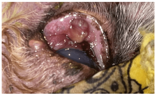
A 14-year-old male poodle dog presented at the clinic with a macroscopic nodule in the palpebral conjunctiva in the upper right eye, measuring 0.7 cm in diameter, white with yellow areas on the cut (Figure 1).
Figure 1. Clinical examination of a skin lesion in a dog
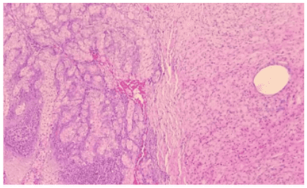
On microscopic examination, benign neoplastic proliferation of epithelial cells was observed, arranged in nests, separated by fine connective tissue trabeculae. The cells had moderately and finely vacuolated cytoplasm, with a round nucleus, loose chromatin and evident nucleolus. In addition, mild anisocytosis, anisokaryosis and multifocal inflammatory infiltrate of lymphocytes and neutrophils were observed. Adjacent to the benign neoplastic proliferation, separated by a layer of connective tissue, a large amount of foamy macrophages arranged in a mantle containing nests of cholesterol slits was also observed (Figure 2).
Figure 2. Histopathological examination of xanthoma
Based on the clinical and laboratory examination, the diagnosis was palpebral conjunctiva: xanthoma. Since it is a benign neoplasm, with good histopathological prognosis upon complete surgical excision.
The presence of xanthoma adjacent to the neoplasm indicates an associated hyperlipidemia. Thus, the patient underwent surgery to excise the lesion using the microsurgical technique.
After pre-anesthetic consultation and physical evaluation, it was found that the patient was fit for the anesthetic procedure. Therefore, the following procedures were performed: Pre-anesthetic medication (PAM) intramuscularly (IM) with acepromazine 0.02 mg/Kg and methadone 0.2 mg/Kg.
Anesthetic induction was performed with propofol 2 mg/Kg, fentanyl 5 mcg/Kg, where the patient allowed intubation and achieved the desired anesthetic plane.
Anesthetic maintenance was performed with remifentanil 10 mg/Kg/hour and isoflurane. Extraconal (peribulbar) locoregional blockade was performed at a dose of 0.1 ml/Kg. The surgical technique started with a quadrangular incision around the lesion and removal was performed. Subsequently, cryotherapy was performed with the CrioFast device.
For post-surgical medication, moxifloxacin QUAD eye drops were prescribed for 15 days.
DISCUSSION
Xanthomas are benign granuloma lesions associated with disrupted lipid transport. They result from abnormalities in the synthesis or degradation of plasma proteins, leading to hyperlipoproteinemia. They are associated with hyperlipoproteinemia and can be caused by the consumption of foods with a high fat content. Alternatively, they can be triggered by diseases such as: diabetes mellitus. In addition, hyperlipoproteinemia may occur in conjunction with an inherited defect in lipid metabolism. The formation of xanthomas is usually secondary to infiltration or deposition of lipoproteins in tissues. Therefore, the original cause of the xanthoma must be part of the clinical analysis so that it can be treated through medication and/or diet (PIRES, 2016).
Hyperlipidemia is characterized as a hereditary disorder of lipoprotein metabolism, with unknown etiology and may be linked to a genetic problem in lipoprotein lipase or the absence of apoprotein CII. Clinical signs usually involve: spontaneous atherosclerosis, retinal lipemia, skin xanthomas, abdominal pain, lethargy, vomiting and/or diarrhea. Neurological manifestations, such as seizures and behavioral changes, can also occur (SALES et al., 2021). Therefore, in the case in question, the investigation of the disease and its treatment become essential.
Eyelid lesions in dogs are relevant in clinical practice, as they have a great impact on the animal’s quality of life, as they cause constant friction with the ocular surfaces (FERREIRA et al., 2009).
As these lesions are usually asymptomatic, animal tutors seek veterinary assistance due to the resemblance of the tumor to neoplastic lesions, however, there are asymptomatic cases that affect organic functions (STILES, 2013).
Clinically, xanthomas are multifocal and consist of papules, nodules or white/yellowish plaques, the edges of which may be erythematous. However, as they are usually associated with hyperlipidemia, it is essential to analyze the presence of signs of this pathology, which are: vomiting, diarrhea and abdominal discomfort and, in more severe cases, when the serum concentration of triglycerides is greater than 1000 mg/dl, may be associated with: pancreatitis, lipemia retinalis, seizures, skin xanthomas, paralysis of peripheral nerves and behavioral changes (STILES, 2013).
For diagnosis, clinical and laboratory tests are used, with biopsy of the lesion for histopathological analysis and definitive diagnosis. Cytology of granulomas can be used as a complementary exam. In addition, because xanthomas are associated with dyslipidemia and secondary diseases, it is important that a biochemical panel be performed, including cholesterol and triglycerides, to analyze or rule out the existence or not of hyperlipidemia and also diagnose diabetes mellitus, for example. If any other pathology is suspected, investigation for diagnosis and treatment should be carried out (PIRES, 2016).
On histopathological examination, xanthomas present diffuse nodular infiltration by macrophages in the connective tissue, with foamy cytoplasm and multinucleated histiocytic giant cells. Macrophages pass through collagen fibers or are in diffuse layers, changing the normal architecture. In addition, in some cases, macrophages and giant cells may be individualized (STILES, 2013).
For treatment, it is essential to assess the etiologic cause of the xanthoma. For cases associated with secondary hyperlipoproteinemia, especially when it is diabetes mellitus, treatment of the cause usually leads to resolution of the xanthoma. In cases of hyperlipidemia, especially if associated with food and idiopathic xanthomatosis, the animal’s diet should be changed to a low-fat diet, which normally leads to improvement in the xanthoma.
With regard to hypertriglyceridemia, adequate treatment is important to avoid future pancreatitis, and treatment to reduce the serum concentration of triglycerides should be performed. In case of chylomicronemia, the diet should be changed with values below 20% of fat or even lower. If the animal is obese, in addition to the restricted fat diet, caloric restriction must be applied. After 6 to 8 weeks on the diet, serum triglyceride and cholesterol levels should be reassessed. Also, drugs for the treatment of hypertriglyceridemia can be administered, but only if serum triglyceride values are greater than 500 mg/dl, niacin 100 mg/day or derivatives of fibric acid (fibrates) such as gemfibrozil 200 mg/day may be indicated for canines and, for cats, 10 mg/kg BID. For hypercholesterolemia, drug treatment should only be performed if serum cholesterol levels are above 800 mg/dl, and a restricted diet should first be attempted (PIRES, 2016).
Ferreira et al. (2009), indicate that the treatment of eyelid tumors in dogs and cats should be done through surgical excision, and the most appropriate technique should be chosen for each case according to the location of the tumor, extension and depth, in addition, whenever If possible, excision with wide surgical margins is preferable to prevent recurrence and always treat the underlying disease before or in conjunction with surgery.
FINAL CONSIDERATIONS
This article aimed to describe a case report of a poodle dog diagnosed with xanthoma, guided by the question: what procedures should be performed for the treatment of xanthoma in a poodle dog? In view of the above, it was found that, in the report presented, the patient evolved clinically favorably and had complete recovery. Thus, it was found that eyelid lesions in dogs are relevant in clinical practice and have a great impact on the animal’s quality of life, as they cause constant friction with the ocular surfaces.
With regard to procedures, the literature points out that, for the treatment of eyelid tumors in dogs and cats, surgical excision must be performed, and the most appropriate technique must be chosen for each case according to the location of the tumor, extension and depth, moreover, whenever possible, excision with wide surgical margins is preferable to prevent recurrence and jointly treat the primary disease, if any.
REFERENCES
FERREIRA, F. M. et al. Neoplasias Oculares In: DALECK, C. R.; DE NARDI, A. B.; RODASKY, S. Oncologia em cães e gatos. São Paulo: Roca, 2009. p.364-384.
PIRES, Andrea do Rosário. Xantoma retrobulbar em gato europeu comum – estudo de um caso clínico. 2016. 100 f. Dissertação (Mestrado em Veterinária) – Universidade de Lisboa. Lisboa, 2016.
SALES, Nathali Adrielli Agassi de et al. Reactive Seizures Due to Hyperlipidemia in a Maltese Dog. Acta Scientiae Veterinariae, [S.L.], v. 49, p. 1, 1 jan. 2021. Disponível em: http://dx.doi.org/10.22456/1679-9216.110100. Acesso em: 26 set. 2022.
STILES, J. Feline Ophthalmology. In: GELLAT, K. N.; GILGER, B. C.; KERN, T. J. (edts). Veterinary ophthalmology. 5ª ed. p 1477-1560. Oxford: John Wiley & Sons, Inc. 2013.
[1] Master’s Degree in Health Sciences from the Universidade Estadual de Ponta Grossa – UEPG; Post-graduation in Ophthalmology and Microsurgery in Veterinary Ophthalmology by Faculdade Qualittas, Graduation in Veterinary Medicine Centro de Ensino Superior dos Campos Gerais – CESCAGE. ORCID: 0000-0003-2979-3094.
[2] Specialization in PAV Anesthesiology, Graduation in Veterinary Medicine by the Centro de Ensino Superior dos Campos Gerais – CESCAGE. ORCID: 0000-0002-4095-3282.
[3] PhD in Animal Pathology; Master in Animal Science with Emphasis in Pathology; Degree in Veterinary Medicine from the Universidade Estadual do Centro Oeste do Paraná – UNICENTRO.
Sent: August, 2022.
Approved: October, 2022.